File:Hilfer1990 Fig08.jpg
From Embryology
Size of this preview: 449 × 599 pixels. Other resolution: 1,498 × 2,000 pixels.
Original file (1,498 × 2,000 pixels, file size: 373 KB, MIME type: image/jpeg)
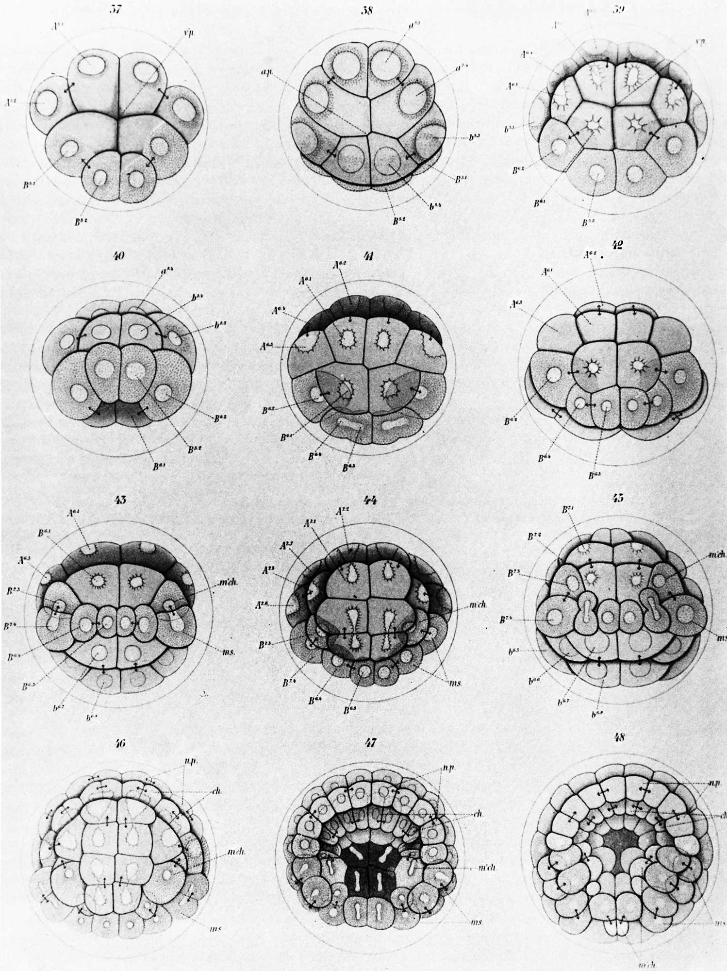
Figure 8. Conklin (1905) early cleavage stages of the tunicate
Diagrams of early cleavage stages of the tunicate, Cynthia partita, from Conklin (1905). The four cells at the bottom of the upper left figure are traced through succeeding cleavages into a series of blastomeres that give rise to segmental muscles and other mesodermal derivatives. This is an example of the painstaking care that was needed to trace regionalization of egg cytoplasm through development.
File history
Yi efo/eka'e gwa ebo wo le nyangagi wuncin ye kamina wunga tinya nan
| Gwalagizhi | Nyangagi | Dimensions | User | Comment | |
|---|---|---|---|---|---|
| current | 10:50, 28 August 2014 | | 1,498 × 2,000 (373 KB) | Z8600021 (talk | contribs) |
You cannot overwrite this file.
File usage
The following page uses this file:


