File:Gray0460.jpg
Original file (818 × 551 pixels, file size: 106 KB, MIME type: image/jpeg)
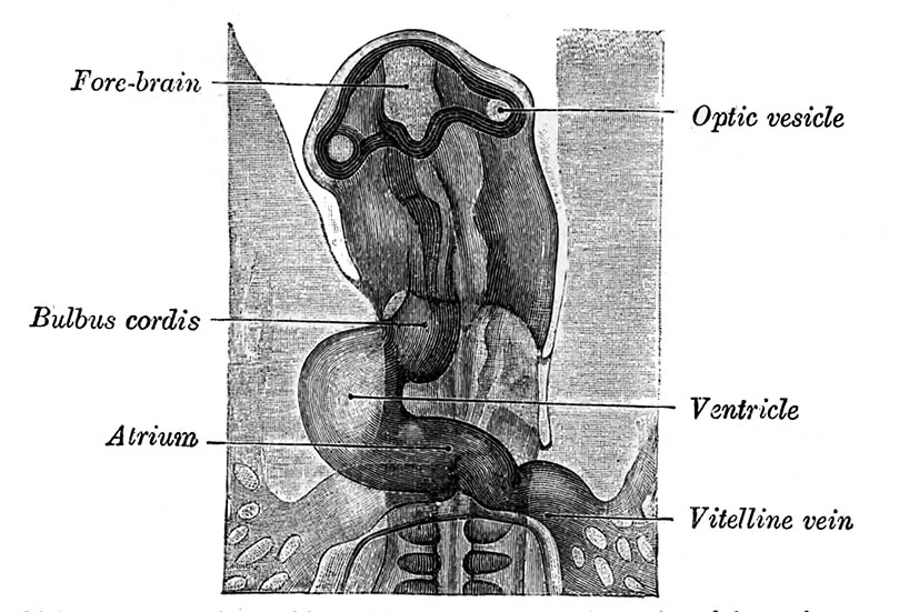
Fig. 460. Head of chick embryo of about thirty-eight hours' incubation
Viewed from the ventral surface. X 26 (Duval.)
(1) the sinus venosus; (2) the primitive atrium; (3) the primitive ventricle; (4) the bulbus cordis, and (5) the truncus arteriosus (Figs. 461, 462). The constriction between the atrium and ventricle constitutes the atrial canal, and indicates the site of the future atrioventricular valves.
| Historic Disclaimer - information about historic embryology pages |
|---|
 |
- Gray's Images: Development | Lymphatic | Neural | Vision | Hearing | Somatosensory | Integumentary | Respiratory | Gastrointestinal | Urogenital | Endocrine | Surface Anatomy | iBook | Historic Disclaimer
| Historic Disclaimer - information about historic embryology pages |
|---|
|
| iBook - Gray's Embryology | |
|---|---|
|
|
Reference
Gray H. Anatomy of the human body. (1918) Philadelphia: Lea & Febiger.
Cite this page: Hill, M.A. (2026, October 3) Embryology Gray0460.jpg. Retrieved from https://embryology.med.unsw.edu.au/embryology/index.php/File:Gray0460.jpg
- © Dr Mark Hill 2026, UNSW Embryology ISBN: 978 0 7334 2609 4 - UNSW CRICOS Provider Code No. 00098G[Category:Chicken]]
File history
Yi efo/eka'e gwa ebo wo le nyangagi wuncin ye kamina wunga tinya nan
| Gwalagizhi | Nyangagi | Dimensions | User | Comment | |
|---|---|---|---|---|---|
| current | 18:14, 23 August 2014 | | 818 × 551 (106 KB) | Z8600021 (talk | contribs) | |
| 18:12, 23 August 2014 | 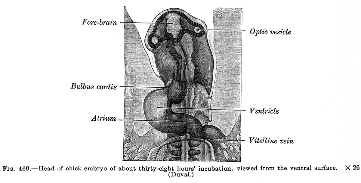 | 1,200 × 608 (124 KB) | Z8600021 (talk | contribs) |
You cannot overwrite this file.
File usage
The following page uses this file:


