File:Embryonic neck muscle cartoon.jpg
Embryonic_neck_muscle_cartoon.jpg (600 × 570 pixels, file size: 45 KB, MIME type: image/jpeg)
Embryonic Neck Muscle Cartoon
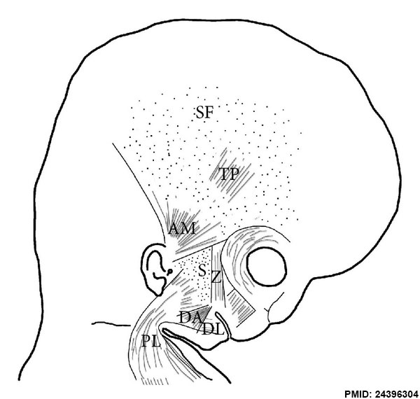
Schematic diagram of the arrangement of the anlage of the superficial musculoaponeurotic system (S). The platysma muscle (PL) derives from the cervical lamina and its mandibular extension. The depressor labii inferioris (DA) and depressor anguli oris (DL) muscles derive from the mandibular lamina. The zygomaticus major muscle (Z) derives from the infraorbital lamina. The superficial layer of the temporal fascia (SF) and the auricularis (AM) and temporoparietalis (TP) muscles originate from the temporal lamina.
Reference
<pubmed>24396304 </pubmed>| ScientificWorldJournal.
Copyright
Copyright © 2013 C. De la Cuadra-Blanco et al. This is an open access article distributed under the Creative Commons Attribution License, which permits unrestricted use, distribution, and reproduction in any medium, provided the original work is properly cited.
Figure 9 716962.fig.009.jpg
ScientificWorldJournal. 2013 Dec 12;2013:716962. doi: 10.1155/2013/716962. eCollection 2013. Development of the platysma muscle and the superficial musculoaponeurotic system (human specimens at 8-17 weeks of development). De la Cuadra-Blanco C1, Peces-Peña MD2, Carvallo-de Moraes LO3, Herrera-Lara ME2, Mérida-Velasco JR1. Author information
Abstract There is controversy regarding the description of the different regions of the face of the superficial musculoaponeurotic system (SMAS) and its relationship with the superficial mimetic muscles. The purpose of this study is to analyze the development of the platysma muscle and the SMAS in human specimens at 8-17 weeks of development using an optical microscope. Furthermore, we propose to study the relationship of the anlage of the SMAS and the neighbouring superficial mimetic muscles. The facial musculature derives from the mesenchyme of the second arch and migrates towards the different regions of the face while forming premuscular laminae. During the 8th week of development, the cervical, infraorbital, mandibular, and temporal laminae are observed to be on the same plane. The platysma muscle derives from the cervical lamina and its mandibular extension enclosing the lower part of the parotid region and the cheek, while the SMAS derives from the upper region. During the period of development analyzed in this study, we have observed no continuity between the anlage of the SMAS and that of the superficial layer of the temporal fascia and the zygomaticus major muscle. Nor have we observed any structure similar to the SMAS in the labial region. PMID 24396304
File history
Yi efo/eka'e gwa ebo wo le nyangagi wuncin ye kamina wunga tinya nan
| Gwalagizhi | Nyangagi | Dimensions | User | Comment | |
|---|---|---|---|---|---|
| current | 11:18, 28 March 2014 | | 600 × 570 (45 KB) | Z8600021 (talk | contribs) | ==Embryonic Neck Muscle Cartoon== Schematic diagram of the arrangement of the anlage of the superficial musculoaponeurotic system (S). The platysma muscle (PL) derives from the cervical lamina and its mandibular extension. The depressor labii inferior... |
You cannot overwrite this file.
File usage
There are no pages that use this file.