File:Mouse CT E10.5 head.jpg
Original file (1,200 × 449 pixels, file size: 83 KB, MIME type: image/jpeg)
Mouse Embryo Computed Tomography
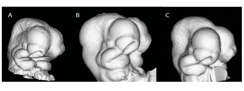
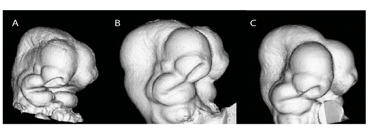
Identically scaled renderings representing 3D μCT scans of embryos (E10.5 head according to paper methods) subjected to different fixation and scanning procedures.
- A - 4% formaldehyde and secondary Bouin's fixation.
- B - 4% formaldehyde + 1% glutaraldehyde with Iothalamate meglumine used as a contrast agent.
- C - 4% formaldehyde + 5% glutaraldehyde plus contrast agent.
All embryos are shown in right 3/4 inferolateral view.
- "Typical μCT renderings are represented by the embryos depicted in Figure 2A-C. The combination of aldehyde fixation and Cysto Conray® yields images (Figure 2B-C) with a surface quality comparable to embryos fixed with Bouin's solution (Figure 2A). Bouin's-fixed embryos appear have a rougher surface texture compared to those fixed with either glutaraldehyde mixture (Figure 2B-C). The identically scaled images of Bouin's fixed embryos appeared generally smaller compared to those fixed with glutaraldehyde mixtures, but a comparison of μCT images only cannot indicate the relative roles of fixation-induced tissue shrinkage or swelling."
Original File name: 1471-213X-10-18-2-l.jpg http://www.biomedcentral.com/1471-213X/10/18/figure/F2
Reference
<pubmed>20163731</pubmed>| BMC Developmental Biology
© 2010 Schmidt et al; licensee BioMed Central Ltd. This is an Open Access article distributed under the terms of the Creative Commons Attribution License (http://creativecommons.org/licenses/by/2.0), which permits unrestricted use, distribution, and reproduction in any medium, provided the original work is properly cited.
File history
Yi efo/eka'e gwa ebo wo le nyangagi wuncin ye kamina wunga tinya nan
| Gwalagizhi | Nyangagi | Dimensions | User | Comment | |
|---|---|---|---|---|---|
| current | 12:24, 17 August 2010 | | 1,200 × 449 (83 KB) | S8600021 (talk | contribs) | ==Mouse Embryo Computed Tomography== Identically scaled renderings representing 3D μCT scans of embryos (E10.5 head according to paper methods) subjected to different fixation and scanning procedures. A, 4% formaldehyde and secondary Bouin's fixation. |
You cannot overwrite this file.
File usage
The following page uses this file: