File:Bailey386.jpg
From Embryology
No higher resolution available.
Bailey386.jpg (491 × 410 pixels, file size: 53 KB, MIME type: image/jpeg)
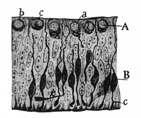
Fig. 386. Section through the wall of the fore-brain vesicle of a chick embryo of 3.5 days
Cajal.
A, b arid c, Differentiating nerve cells in apolar stage, the neurofibrils are black; a, cell in a stage transitional to the bipolar stage; B, bipolar cells; c (at lower right corner), cone of "growth" of developing axone; e, tangential axone. The cells in the bipolar stage have migrated out ward, but the neuroblast or mantle layer has not yet been differentiated.
File history
Yi efo/eka'e gwa ebo wo le nyangagi wuncin ye kamina wunga tinya nan
| Gwalagizhi | Nyangagi | Dimensions | User | Comment | |
|---|---|---|---|---|---|
| current | 01:36, 30 Ocak 2011 | | 491 × 410 (53 KB) | S8600021 (talk | contribs) | ==Fig. 386. Section through the wall of the fore-brain vesicle of a chick embryo of 3.5 days== Cajal. A, b arid c, Differentiating nerve cells in apolar stage, the neurofibrils are black; a, cell in a stage transitional to the bipolar stage; B, bipolar |
You cannot overwrite this file.
File usage
The following 4 pages use this file:


