Category:Blue Histology
From Embryology
This Embryology category relates to Blue Histology images copyright Lutz Slomianka 1998-2009.
The literary and artistic works on the original Blue Histology website may be reproduced, adapted, published and distributed for non-commercial purposes.
Pages in category 'Blue Histology'
The following 5 pages are in this category, out of 5 total.
Media in category 'Blue Histology'
The following 200 files are in this category, out of 431 total.
(previous page) (next page)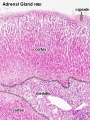 Adrenal histology 001.jpg 450 × 600; 151 KB
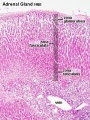 Adrenal histology 002.jpg 450 × 600; 150 KB
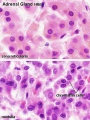 Adrenal histology 003.jpg 450 × 600; 71 KB
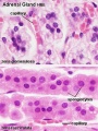 Adrenal histology 004.jpg 450 × 600; 71 KB
 Adrenal histology 005.jpg 1,280 × 1,024; 298 KB
 Adrenal histology 006.jpg 1,280 × 1,024; 313 KB
 Adrenal histology 007.jpg 1,280 × 1,024; 298 KB
 Adrenal histology 008.jpg 1,280 × 1,024; 272 KB
 Adrenal histology 009.jpg 1,280 × 1,024; 265 KB
 Adrenal histology 010.jpg 1,280 × 1,024; 292 KB
 Adrenal histology 011.jpg 1,280 × 1,024; 572 KB
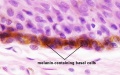
 Adult epidermis histology 01.jpg 600 × 750; 83 KB
 Adult epidermis histology 02.jpg 600 × 750; 123 KB
 Adult epidermis histology 03.jpg 600 × 375; 46 KB
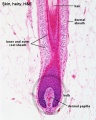
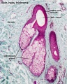
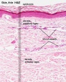 Adult skin histology 01.jpg 600 × 750; 73 KB
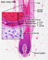
 Adult skin histology 02.jpg 600 × 750; 88 KB
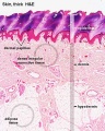
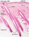
 Adult skin histology 03.jpg 600 × 750; 108 KB
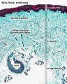
 Adult skin histology 04.jpg 480 × 600; 128 KB
 Apocrine secretion animation.gif 60 × 80; 4 KB
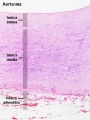
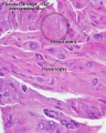
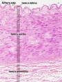 Artery histology 01.jpg 400 × 533; 80 KB
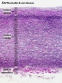
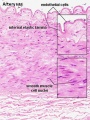 Artery histology 02.jpg 400 × 533; 78 KB
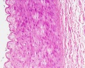
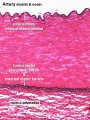 Artery histology 03.jpg 400 × 533; 89 KB
 Artery histology 04.jpg 800 × 1,000; 93 KB
 Artery histology 05.jpg 400 × 533; 72 KB
 Artery histology 06.jpg 400 × 533; 91 KB
 Artery histology 11.jpg 1,280 × 1,024; 344 KB
 Artery histology 12.jpg 1,280 × 1,024; 206 KB
 Artery histology 13.jpg 1,280 × 1,024; 474 KB
 Artery histology 14.jpg 1,280 × 1,024; 466 KB
 Artery histology 15.jpg 1,280 × 1,024; 343 KB
 Artery histology 16.jpg 1,280 × 1,024; 409 KB
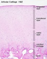
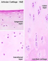 Articular cartilage 01.jpg 500 × 626; 42 KB
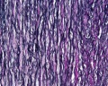
 Articular cartilage.jpg 500 × 626; 77 KB
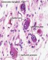 Autonomic ganglion histology 01.jpg 641 × 800; 56 KB
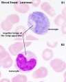
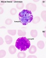 Basophil 01.jpg 480 × 600; 37 KB
 Bladder histology 001.jpg 1,280 × 1,024; 522 KB
 Bladder histology 002.jpg 1,280 × 1,024; 295 KB
 Bladder histology 003.jpg 1,280 × 1,024; 229 KB
 Bladder histology 004.jpg 1,280 × 1,024; 212 KB
 Bladder histology 01.jpg 480 × 600; 29 KB
 Bladder histology.jpg 300 × 400; 56 KB
 Blood vessel wall cartoon.jpg 450 × 600; 71 KB
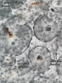
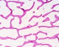
 Bone histology 001.jpg 1,280 × 1,024; 276 KB
 Bone histology 002.jpg 1,280 × 1,024; 309 KB
 Bone histology 003.jpg 1,280 × 1,024; 663 KB
 Bone histology 004.jpg 1,280 × 1,024; 605 KB
 Bone histology 005.jpg 1,280 × 1,024; 529 KB
 Bone histology 006.jpg 1,280 × 1,024; 360 KB
 Bone histology 007.jpg 1,280 × 1,024; 299 KB
 Bone histology 008.jpg 1,280 × 1,024; 550 KB
 Bone histology 009.jpg 1,280 × 1,024; 444 KB
 Bone histology 010.jpg 1,280 × 1,024; 256 KB
 Bone histology 011.jpg 1,280 × 1,024; 348 KB
 Bone histology 012.jpg 1,280 × 1,024; 165 KB
 Bone histology 013.jpg 1,280 × 1,024; 210 KB
 Bone histology 014.jpg 1,280 × 1,024; 541 KB
 Bone histology 015.jpg 1,280 × 1,024; 519 KB
 Bone histology 016.jpg 1,280 × 1,024; 379 KB
 Bone histology 017.jpg 1,280 × 1,024; 442 KB
 Bone histology 018.jpg 1,280 × 1,024; 336 KB
 Bone histology 019.jpg 1,280 × 1,024; 275 KB
 Bone histology 020.jpg 1,280 × 1,024; 272 KB
 Bone histology 021.jpg 1,280 × 1,024; 254 KB
 Bone histology 022.jpg 2,500 × 2,000; 328 KB
 Bone histology 066.jpg 2,500 × 2,000; 361 KB
 Bone histology 101.jpg 400 × 533; 59 KB
 Bone histology 111.jpg 400 × 533; 70 KB
 Bone histology 112.jpg 400 × 533; 46 KB
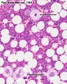
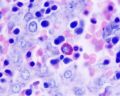
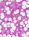 Bone marrow histology 01.jpg 480 × 600; 114 KB
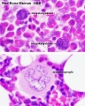
 Bone marrow histology 02.jpg 480 × 600; 109 KB
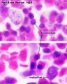
 Bone marrow histology 03.jpg 480 × 600; 81 KB
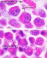
 Bone marrow histology 04.jpg 480 × 600; 61 KB
 Bone marrow histology 05.jpg 480 × 600; 62 KB
 Bone-bon02he.jpg 1,280 × 1,024; 348 KB
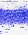
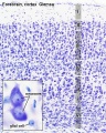 Brain histology 01.jpg 480 × 600; 125 KB
 Brain histology 02.jpg 480 × 600; 51 KB
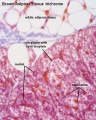 Brown adipose histology.jpg 400 × 500; 64 KB
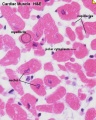
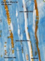 Cardiac muscle histology.jpg 300 × 400; 42 KB
 Cartilage em01.jpg 800 × 551; 176 KB
 Cartilage histology 001.jpg 1,280 × 1,024; 362 KB
 Cartilage histology 002.jpg 1,280 × 1,024; 174 KB
 Cartilage histology 003.jpg 1,280 × 1,024; 161 KB
 Cartilage histology 004.jpg 1,280 × 1,024; 216 KB
 Cartilage histology 005.jpg 1,280 × 1,024; 344 KB
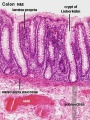
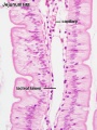
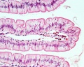
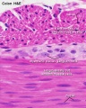
 Colon histology 001.jpg 400 × 533; 64 KB
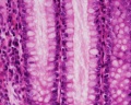
 Colon histology 002.jpg 300 × 400; 73 KB
 Colon histology 003.jpg 1,280 × 1,024; 117 KB
 Colon histology 004.jpg 1,280 × 1,024; 292 KB
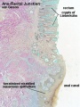 Colon histology 006.jpg 400 × 533; 70 KB
 Colon histology 007.jpg 1,280 × 1,024; 152 KB
 Colon histology 008.jpg 1,278 × 959; 237 KB
 Colon histology 009.jpg 1,280 × 1,024; 159 KB
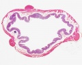
 Colon MALT.jpg 500 × 333; 67 KB
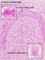 Corpus luteum lutein cells.jpg 450 × 600; 104 KB
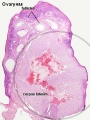 Corpus luteum.jpg 450 × 600; 94 KB
 Dorsal root ganglion histology 01.jpg 640 × 800; 45 KB
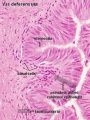
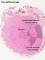 Ductus deferens 01.jpg 400 × 533; 76 KB
 Ductus deferens 02.jpg 400 × 533; 80 KB
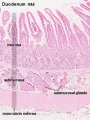
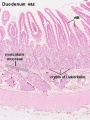
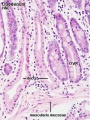
 Duodenum histology 01.jpg 480 × 600; 83 KB
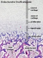
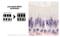
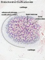 Endochondral ossification 1.jpg 400 × 534; 91 KB
 Endochondral ossification 2.jpg 400 × 533; 99 KB
 Endochondral ossification.jpg 400 × 533; 91 KB
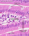
 Epididymis histology 01.jpg 600 × 375; 20 KB
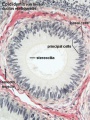 Epididymis histology 03.jpg 400 × 533; 68 KB
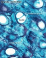
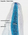 Epiglottis cartilage 01.jpg 500 × 626; 95 KB
 Epiglottis cartilage 02.jpg 500 × 626; 101 KB
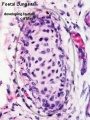
 Fetal cartilage 01.jpg 639 × 400; 71 KB
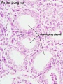
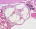
 Fetal lung histology 01.jpg 1,280 × 1,024; 339 KB
 Fetal lung histology 02.jpg 450 × 600; 74 KB
 Fetal lung histology.jpg 450 × 600; 83 KB
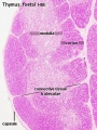 Fetal thymus.jpg 450 × 600; 122 KB
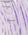 Fibrous cartilage 01.jpg 500 × 626; 76 KB
 Fibrous cartilage 02.jpg 500 × 626; 96 KB
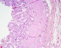
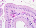
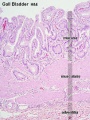 Gall bladder histology 001.jpg 375 × 500; 78 KB
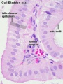 Gall bladder histology 002.jpg 375 × 500; 45 KB
 Gall bladder histology 003.jpg 1,280 × 1,024; 577 KB
 Gall bladder histology 004.jpg 1,280 × 1,024; 254 KB
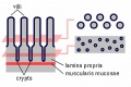
 Gastrointestinal villi and crypts cartoon.jpg 500 × 333; 28 KB
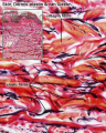
 Hair histology.jpg 600 × 451; 131 KB
 Heart histology 001.jpg 400 × 500; 83 KB
 Heart histology 002.jpg 400 × 500; 81 KB
 Heart histology 003.jpg 400 × 500; 136 KB
 Heart histology 004.jpg 400 × 500; 97 KB
 Heart histology 101.jpg 1,280 × 1,024; 258 KB
 Heart histology 102.jpg 1,280 × 1,024; 242 KB
 Heart histology 103.jpg 1,280 × 1,024; 281 KB
 Heart histology 104.jpg 1,280 × 1,024; 280 KB
 Heart histology 105.jpg 1,280 × 1,024; 379 KB
 Heart histology 106.jpg 1,280 × 1,024; 347 KB
 Heart histology 107.jpg 1,280 × 1,024; 395 KB
 Heart-histology-102.jpg 1,280 × 1,024; 242 KB
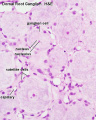
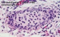
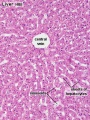
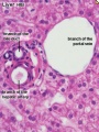
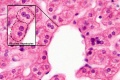
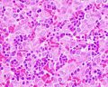
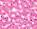
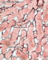
 Histology-fetal liver HEx100.jpg 1,280 × 1,024; 214 KB
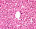
 Histology-fetal liver HEx40.jpg 1,000 × 800; 281 KB
 Holocrine secretion animation.gif 60 × 80; 16 KB
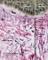
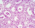
 Human fetal kidney histology 01.jpg 1,280 × 1,024; 481 KB
 Human fetal kidney histology 02.jpg 1,280 × 1,024; 322 KB
 Human fetal kidney histology 03.jpg 1,280 × 1,024; 333 KB
 Human fetal kidney histology 04.jpg 1,280 × 1,024; 307 KB
 Hyaline cartilage 01.jpg 500 × 626; 71 KB
 Hyaline cartilage 02.jpg 500 × 626; 62 KB
 Hyaline cartilage 03.jpg 500 × 626; 92 KB
 Hyaline cartilage 04.jpg 500 × 626; 101 KB
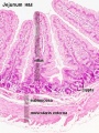
 Ileum histology 01.jpg 480 × 600; 69 KB
 Integumentary histology 01.jpg 480 × 600; 70 KB
 Integumentary histology 04.jpg 280 × 700; 65 KB
 Integumentary histology 10.jpg 800 × 1,000; 101 KB
 Integumentary- hair follicle 01.jpg 479 × 599; 66 KB
 Integumentary- hair follicle 02.jpg 479 × 599; 74 KB
 Integumentary- hair follicle 03.jpg 479 × 599; 48 KB
 Integumentary- sebaceous gland histology 01.jpg 400 × 500; 125 KB
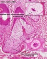 Integumentary- sebaceous gland histology 02.jpg 400 × 500; 150 KB
 Intestine histology 001.jpg 450 × 600; 65 KB
 Intestine histology 002.jpg 800 × 640; 130 KB
 Intestine histology 003.jpg 400 × 533; 64 KB
 Intestine histology 004.jpg 400 × 533; 81 KB
 Intestine histology 005.jpg 400 × 533; 78 KB
 Intestine histology 006.jpg 400 × 533; 77 KB
 Intestine histology 007.jpg 400 × 533; 82 KB
 Intestine villi crypts cartoon.jpg 500 × 334; 29 KB
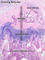
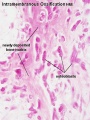 Intramembranous ossification centre.jpg 450 × 600; 69 KB
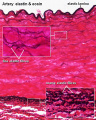
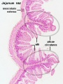
 Jejunum histology 01.jpg 480 × 600; 79 KB
 Liver animated cartoon.gif 300 × 200; 239 KB
 Liver histology 001.jpg 400 × 533; 94 KB
 Liver histology 002.jpg 375 × 500; 54 KB
 Liver histology 003.jpg 375 × 500; 52 KB
 Liver histology 004.jpg 600 × 400; 70 KB
 Liver histology 008.jpg 1,280 × 1,024; 214 KB
 Liver histology 009.jpg 1,280 × 1,024; 373 KB
 Liver histology 101.jpg 1,280 × 1,024; 410 KB
 Liver histology 102.jpg 1,280 × 1,024; 475 KB
 Liver histology 103.jpg 1,280 × 1,024; 330 KB
 Liver histology EM01.jpg 1,028 × 708; 141 KB
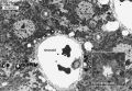 Liver histology EM02.jpg 1,028 × 707; 154 KB
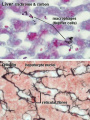 Liver- Kupffer cell and reticular fibre.jpg 600 × 800; 49 KB
 Liver-reticular fibre.jpg 700 × 875; 77 KB
 Lymph node 05.jpg 1,000 × 800; 180 KB
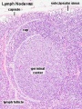
 Lymph node histology 01.jpg 600 × 400; 61 KB
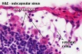
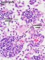
 Lymph node histology 02.jpg 450 × 600; 130 KB
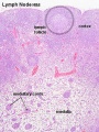
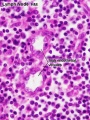
 Lymph node histology 03.jpg 450 × 600; 140 KB
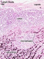
 Lymph node histology 04.jpg 450 × 600; 88 KB
 Lymph node histology 05.jpg 450 × 600; 87 KB
 Lymph node histology 06.jpg 450 × 600; 141 KB
 Lymphocyte 02.jpg 500 × 334; 27 KB
 Merocrine secretion animation.gif 60 × 80; 10 KB
 Mesentery histology 01.jpg 1,280 × 1,024; 139 KB
 Mesentery histology 02.jpg 1,280 × 1,024; 256 KB
 Monkey- ovary primordial follicle.jpg 1,000 × 800; 292 KB
 Monocyte 01.jpg 480 × 600; 39 KB
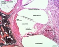
 Mouse organ of corti 01.jpg 1,280 × 1,024; 339 KB
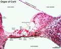
 Mouse organ of corti 02.jpg 1,280 × 1,024; 320 KB
 Mouse organ of corti 03.jpg 1,280 × 1,024; 207 KB
 Mouse organ of corti 04.jpg 1,280 × 1,024; 202 KB
 Mouse organ of corti 05.jpg 1,280 × 1,024; 171 KB
 Muscle fiber types.jpg 400 × 250; 49 KB
 Myelination animation.gif 300 × 200; 77 KB
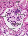 Nephron histology 01.jpg 400 × 500; 79 KB
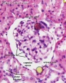 Nephron histology 02.jpg 400 × 500; 77 KB
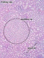 Nephron histology 03.jpg 375 × 500; 97 KB
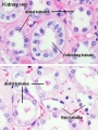 Nephron histology 04.jpg 375 × 500; 54 KB
 Nephron histology.jpg 400 × 500; 70 KB


