File:Lymphatic vessel formation model.jpg
Original file (600 × 692 pixels, file size: 179 KB, MIME type: image/jpeg)
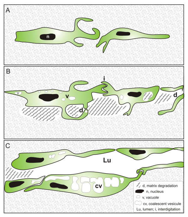
Tunneling model of lymphatic vessel formation
The model is based on ultrastructural observations performed in in vitro and in vivo models of lymphangiogenesis. (A): LEC alignment. Elongated LEC migrate and extend long cytoplasmic protrusions. (B): Vacuolization and matrix degradation. The continuity of LEC lining is mediated by interdigitations (i). Vesicle invaginations lead to the formation of intracellular vacuoles (v) in the cytoplasm and in protrusions. Matrix degradation (d) occurs intracellularly and extracellularly generating space between cells. (C): Luminogenesis. The lumen (lu) is formed de novo in the intercellular space. The intracellular vacuoles coalesce (cv) and likely fuse with the cytoplasmic membrane to increase the lumen.
Reference
Detry et al. BMC Cell Biology 2011 12:29 doi:10.1186/1471-2121-12-29
Original file name: 1471-2121-12-29-8.jpg
File history
Yi efo/eka'e gwa ebo wo le nyangagi wuncin ye kamina wunga tinya nan
| Gwalagizhi | Nyangagi | Dimensions | User | Comment | |
|---|---|---|---|---|---|
| current | 09:55, 5 February 2012 | | 600 × 692 (179 KB) | S8600021 (talk | contribs) | 1471-2121-12-29-8.jpg |
You cannot overwrite this file.
File usage
The following page uses this file: