File:Cat oocyte calcium concentration.jpg
Cat_oocyte_calcium_concentration.jpg (600 × 486 pixels, file size: 80 KB, MIME type: image/jpeg)
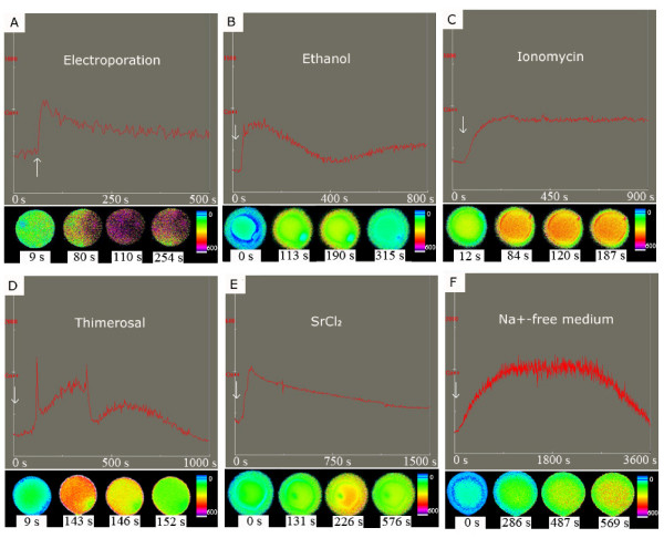
Changes in the intracellular calcium ion concentration of cat oocytes induced by different stimuli
intracellular calcium ion concentration [Ca2+ ]i
The upper part of each section shows cytoplasmic free Ca2+ levels as detected by a photometer; the arrows indicate the time when the stimulus was applied. In the lower part of each section images of [Ca2+ ]i changes in individual cat oocytes are shown; different colors indicate different intracellular free Ca2+ concentrations.
- Links: Cat Development | Oocyte Development
1477-7827-7-148-1.jpg
Reference
http://www.rbej.com/content/7/1/148
Wang et al. Reproductive Biology and Endocrinology 2009 7:148 doi:10.1186/1477-7827-7-148
© 2009 Wang et al; licensee BioMed Central Ltd.
This is an Open Access article distributed under the terms of the Creative Commons Attribution License (http://creativecommons.org/licenses/by/2.0), which permits unrestricted use, distribution, and reproduction in any medium, provided the original work is properly cited.
File history
Click on a date/time to view the file as it appeared at that time.
| Date/Time | Thumbnail | Dimensions | User | Comment | |
|---|---|---|---|---|---|
| current | 12:21, 4 November 2011 |  | 600 × 486 (80 KB) | S8600021 (talk | contribs) | ==Changes in the intracellular calcium ion concentration of cat oocytes induced by different stimuli== intracellular calcium ion concentration [Ca<sup>2+</sup> ]<sub>i</sub> The upper part of each section shows cytoplasmic free Ca2+ levels as detected |
You cannot overwrite this file.
File usage
The following page uses this file:
