File:Kollmann407-409.jpg
Original file (729 × 1,000 pixels, file size: 134 KB, MIME type: image/jpeg)
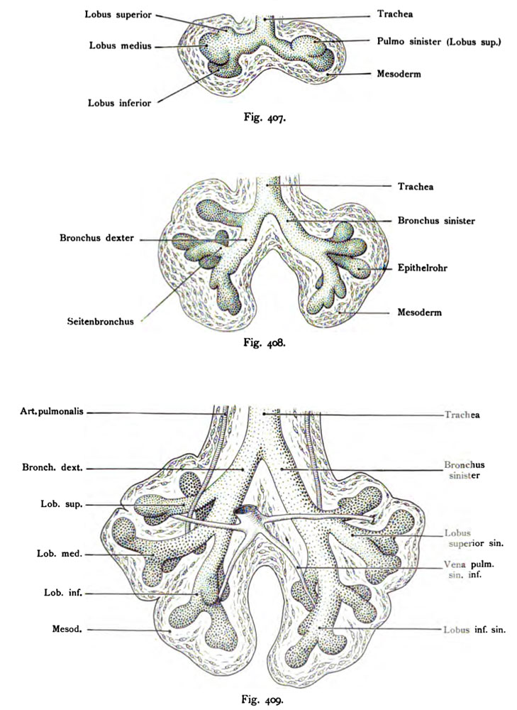
Fig. 407. Pulmonary system of a human embryo, 4.3 mm CRL
Magnified 50 times
(After His.)
The unsymmetrical anlage (primordia) is obvious, the right pulmonary system
is considerably longer than the left. The first traces of
Breakdown shows the left lung sacs two, left three bud-like
Distension. The existing pipe is made of endodermal cells in the interior, including
its branches dotted the surrounding mesoderm shows the known cells of this tissue.
- This text is a Google translate computer generated translation and may contain many errors.
Images from - Atlas of the Development of Man (Volume 2)
(Handatlas der entwicklungsgeschichte des menschen)
- Kollmann Atlas 2: Gastrointestinal | Respiratory | Urogenital | Cardiovascular | Neural | Integumentary | Smell | Vision | Hearing | Kollmann Atlas 1 | Kollmann Atlas 2 | Julius Kollmann
- Links: Julius Kollman | Atlas Vol.1 | Atlas Vol.2 | Embryology History
| Historic Disclaimer - information about historic embryology pages |
|---|
 |
Reference
Kollmann JKE. Atlas of the Development of Man (Handatlas der entwicklungsgeschichte des menschen). (1907) Vol.1 and Vol. 2. Jena, Gustav Fischer. (1898).
Cite this page: Hill, M.A. (2026, October 4) Embryology Kollmann407-409.jpg. Retrieved from https://embryology.med.unsw.edu.au/embryology/index.php/File:Kollmann407-409.jpg
- © Dr Mark Hill 2026, UNSW Embryology ISBN: 978 0 7334 2609 4 - UNSW CRICOS Provider Code No. 00098G
Fig. 407. Lungenanlage eines menschlichen Embryo,
4,3 mm Nackensteißlänge. 50 mal vergr.
(Nach His.)
Die unsymmetrische Anlage ist sehr auflfallend, die rechte Lungenanlage ist erheblich länger als die linke und zugleich weiter. Als erste Spuren der Gliederung zeigt das linke Lungensäckchen zwei, das linke drei knospenartige Auftreibungen. Das aus Entodermzellen bestehende Rohr im Innern ist samt seinen Verzweigungen punktiert, das umgebende Mesoderm zeigt die bekannten Zellen dieses Gewebes.
File history
Yi efo/eka'e gwa ebo wo le nyangagi wuncin ye kamina wunga tinya nan
| Gwalagizhi | Nyangagi | Dimensions | User | Comment | |
|---|---|---|---|---|---|
| current | 15:18, 16 October 2011 | | 729 × 1,000 (134 KB) | S8600021 (talk | contribs) | {{Kollmann1907}} |
You cannot overwrite this file.
File usage
The following 2 pages use this file:


