File:Mouse lung development 01a.jpg
Original file (800 × 1,003 pixels, file size: 495 KB, MIME type: image/jpeg)
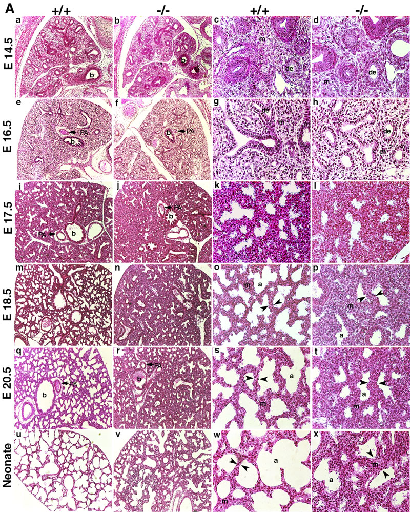
Histological analyses of Mouse Lungs at Various Embryonic Stages
indicates lung sections stained with H and E, and taken at various gestational stages as indicated. The first two columns are at lower magnification (10X) and the last two columns at higher magnification (40X). The first and third columns are representative sections from wild-type (+/+) embryos and the second and fourth columns from knockout (-/-) embryos. No differences were visible at E14.5 or E16.5 (panel A, a-h). Significant differences were visible from E17.5 onwards (panel A, i-x). Distal tubules showed dilation and mesenchyme thinning, with progression of septation in wild-type lungs (panel A, i, m and q), whereas the knockout lungs showed less saccular structures and more mesenchyme (panel A, j, n and r). At higher magnifications of wild-type lungs (panel A, k, o and s), developing pre-alveoli and thinning mesenchyme (arrowheads) were seen but sections from knockout embryos showed a delay in this sequence of development (panel A, l, p and t). At birth, lungs from knockout pups showed deficient septation and thick-walled mesenchyme (panel A, x, arrowheads). Labels: de, distal epithelium; m, mesenchyme; PA, pulmonary artery; a, pre-alveoli; b, bronchi. Panel B shows the morphometric analyses of lung terminal sac spaces in lungs at various gestational stages. A significant reduction in the terminal sac space was noted from E17.5 onwards. Results are expressed as mean ± s.d.
Original file name: 1471-213X-4-1-3.jpg http://www.biomedcentral.com/1471-213X/4/1/figure/F3 (Panel A cropped from original full image, resized to fit screen 800px)
Mouse lung development 01.jpg
Reference
<pubmed>15005800</pubmed>| BMC Developmental Biology
Yu et al. BMC Developmental Biology 2004 4:1 doi:10.1186/1471-213X-4-1
© 2004 Yu et al; licensee BioMed Central Ltd. This is an Open Access article: verbatim copying and redistribution of this article are permitted in all media for any purpose, provided this notice is preserved along with the article's original URL.
File history
Yi efo/eka'e gwa ebo wo le nyangagi wuncin ye kamina wunga tinya nan
| Gwalagizhi | Nyangagi | Dimensions | User | Comment | |
|---|---|---|---|---|---|
| current | 14:52, 25 August 2011 | | 800 × 1,003 (495 KB) | S8600021 (talk | contribs) | ==Histological analyses of Mouse Lungs at Various Embryonic Stages== indicates lung sections stained with H and E, and taken at various gestational stages as indicated. The first two columns are at lower magnification (10X) and the last two columns at hi |
You cannot overwrite this file.
File usage
There are no pages that use this file.