File:Model of human pancreatic islet.jpg
Original file (482 × 702 pixels, file size: 237 KB, MIME type: image/jpeg)
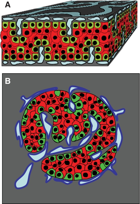
Model of endocrine cell and vessel organization in human islets
A: α-Cells (green) and β-cells (red) are organized into a thick folded plate lined at both sides with vessels (blue). α-Cells are mostly at the periphery of the plate and in close contact with vessels. β-Cells occupy a more central part of the plate and most of them develop cytoplasmic extension that runs between α-cells and reaches the surface of vessels.
B: The plate with adjacent vessels is folded so that it forms an islet.
Original file name: FIG. 7. Zdb0051061080007.jpg http://www.ncbi.nlm.nih.gov/pmc/articles/PMC2857900/figure/F7/
Reference
<pubmed>20185817</pubmed>| PMC2857900 | Diabetes.
© 2010 by the American Diabetes Association
Readers may use this article as long as the work is properly cited, the use is educational and not for profit, and the work is not altered. See http://creativecommons.org/licenses/by-nc-nd/3.0/ for details.
File history
Yi efo/eka'e gwa ebo wo le nyangagi wuncin ye kamina wunga tinya nan
| Gwalagizhi | Nyangagi | Dimensions | User | Comment | |
|---|---|---|---|---|---|
| current | 14:30, 8 August 2011 | | 482 × 702 (237 KB) | S8600021 (talk | contribs) | ==Model of endocrine cell and vessel organization in human islets== A: α-Cells (green) and β-cells (red) are organized into a thick folded plate lined at both sides with vessels (blue). α-Cells are mostly at the periphery of the plate and in close con |
You cannot overwrite this file.
File usage
The following 2 pages use this file:


