File:Model of human pancreatic islet.jpg
Original file (482 × 702 pixels, file size: 237 KB, MIME type: image/jpeg)
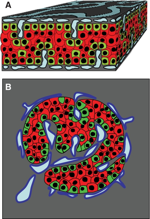
Model of endocrine cell and vessel organization in human islets
A: α-Cells (green) and β-cells (red) are organized into a thick folded plate lined at both sides with vessels (blue). α-Cells are mostly at the periphery of the plate and in close contact with vessels. β-Cells occupy a more central part of the plate and most of them develop cytoplasmic extension that runs between α-cells and reaches the surface of vessels.
B: The plate with adjacent vessels is folded so that it forms an islet.
Original file name: FIG. 7. Zdb0051061080007.jpg http://www.ncbi.nlm.nih.gov/pmc/articles/PMC2857900/figure/F7/
Reference
<pubmed>20185817</pubmed>| PMC2857900 | Diabetes.
Readers may use this article as long as the work is properly cited, the use is educational and not for profit, and the work is not altered. See http://creativecommons.org/licenses/by-nc-nd/3.0/ for details.
File history
Yi efo/eka'e gwa ebo wo le nyangagi wuncin ye kamina wunga tinya nan
| Gwalagizhi | Nyangagi | Dimensions | User | Comment | |
|---|---|---|---|---|---|
| current | 14:30, 8 August 2011 | | 482 × 702 (237 KB) | S8600021 (talk | contribs) | ==Model of endocrine cell and vessel organization in human islets== A: α-Cells (green) and β-cells (red) are organized into a thick folded plate lined at both sides with vessels (blue). α-Cells are mostly at the periphery of the plate and in close con |
You cannot overwrite this file.
File usage
The following 2 pages use this file:


