File:Bradley1904a plate49.jpg
Original file (1,280 × 1,743 pixels, file size: 302 KB, MIME type: image/jpeg)
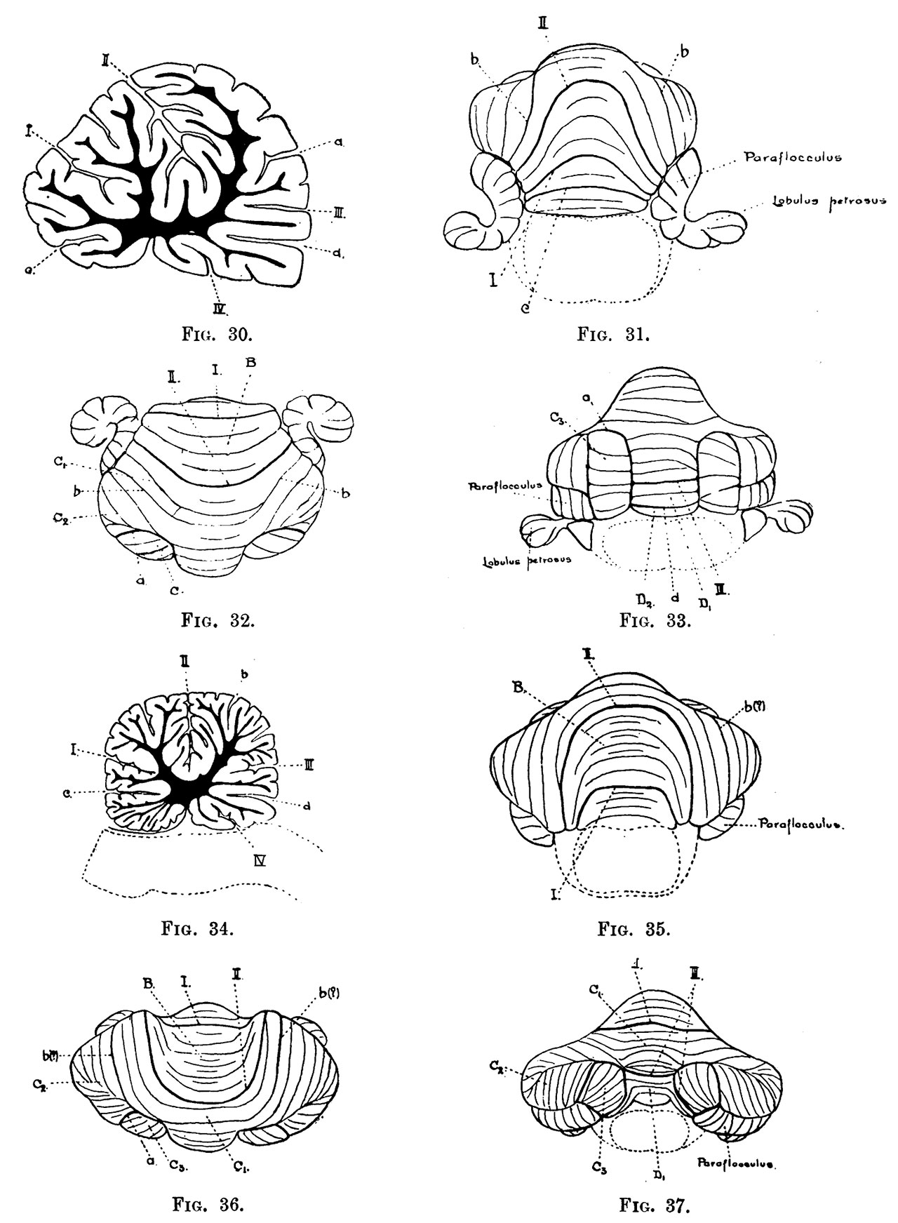
Plate 49
Fig. 27. Didelphis azare. Superior view. x 2.
Fig. 28. Didelphis azare. x 2. Anterior view. x 2.
Fig. 29. Didelphis azare. Postero-lateral view. x 2.
Fig. 30. Didelphis azare. Mesial sagittal section.
Fig. 31. Herpestes mungo. Anterior view. x 2.
Fig. 32. Superior view. x2.
Fig. 33. Posterior view. x 2.
Fig. 34. Mesial sagittal section.
” . Procyon lotor. Anterior view. x1.
” Superior view. x1, ” Posterior view. x1. Mesial sagittal section.’
” . Viverra civetta. Anterior view. x 1.
» Superior view. x1. ” Posterior view. x1. The dotted areas are connected with both lobe C and lobule D,. Mesial sagittal section.
” . Viverra malaccensis. Posterior view. x 1. . Phoca vitulina. Superior view. x 4.
» Posterior view. x 4. ” Inferior view. x }.
Reference
Bradley OC. The mammalian cerebellum: its lobes and fissures. (1904) J Anat Physiol. 38(4): 448-475. PMID17232617
Cite this page: Hill, M.A. (2026, October 4) Embryology Bradley1904a plate49.jpg. Retrieved from https://embryology.med.unsw.edu.au/embryology/index.php/File:Bradley1904a_plate49.jpg
- © Dr Mark Hill 2026, UNSW Embryology ISBN: 978 0 7334 2609 4 - UNSW CRICOS Provider Code No. 00098G
File history
Yi efo/eka'e gwa ebo wo le nyangagi wuncin ye kamina wunga tinya nan
| Gwalagizhi | Nyangagi | Dimensions | User | Comment | |
|---|---|---|---|---|---|
| current | 19:43, 14 Mayıs 2020 | | 1,280 × 1,743 (302 KB) | Z8600021 (talk | contribs) | |
| 18:31, 14 Mayıs 2020 |  | 1,607 × 2,478 (428 KB) | Z8600021 (talk | contribs) | ==Plate 49== ===Reference=== {{Ref-Bradley1904a}} {{footer}} |
You cannot overwrite this file.
File usage
The following 2 pages use this file:


