File:Nephron EM11.jpg
Original file (2,983 × 2,000 pixels, file size: 398 KB, MIME type: image/jpeg)
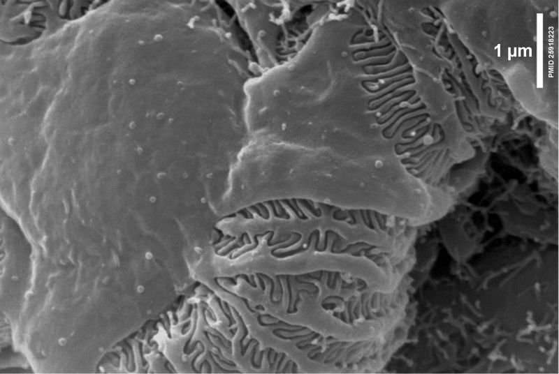
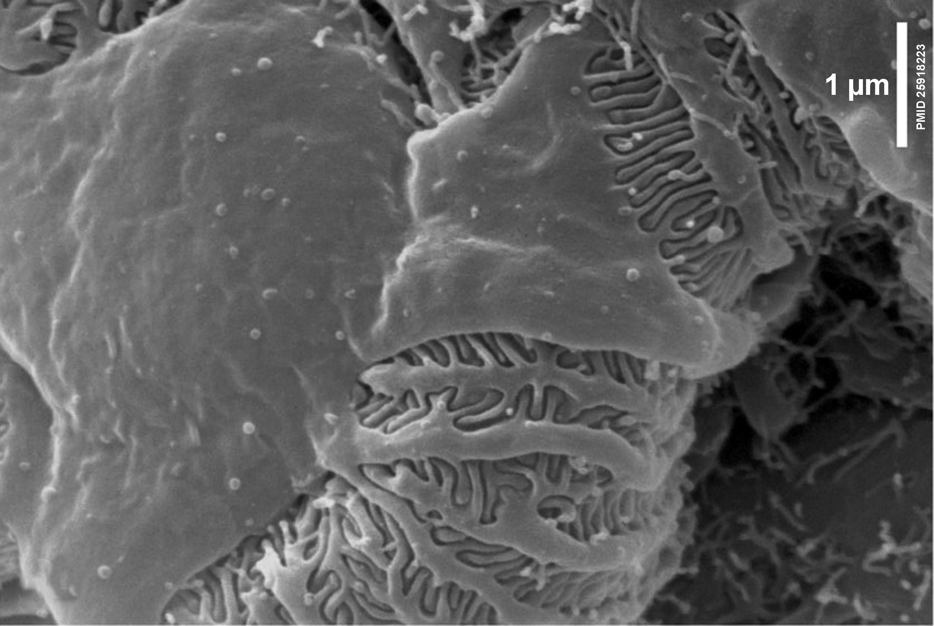
Glomerulus Podocyte Electron Micreograph
Electron Micrograph of the nephron glomerulus showing podocyte and glomerular capillary.
This high magnification view shows a podocyte with the interdigitated foot processes (pedicels) that are wrapped around the exterior of a glomerular capillary forming slit diaphragms (SDs).
Scale Bar: 1 µm.
Glomerular filtration barrier - formed by podocytes and the fenestrated endothelium.
- Links: colour image version | renal histology
Reference
Scott RP & Quaggin SE. (2015). Review series: The cell biology of renal filtration. J. Cell Biol. , 209, 199-210. PMID: 25918223 DOI.
Copyright
Rockefeller University Press - Copyright Policy This article is distributed under the terms of an Attribution–Noncommercial–Share Alike–No Mirror Sites license for the first six months after the publication date (see http://www.jcb.org/misc/terms.shtml). After six months it is available under a Creative Commons License (Attribution–Noncommercial–Share Alike 4.0 Unported license, as described at https://creativecommons.org/licenses/by-nc-sa/4.0/ ). (More? Help:Copyright Tutorial)
Figure 2. Panel B cropped resized, relabelled and converted to black and white image. Text above modified from figure legend.
Cite this page: Hill, M.A. (2026, October 2) Embryology Nephron EM11.jpg. Retrieved from https://embryology.med.unsw.edu.au/embryology/index.php/File:Nephron_EM11.jpg
- © Dr Mark Hill 2026, UNSW Embryology ISBN: 978 0 7334 2609 4 - UNSW CRICOS Provider Code No. 00098G
File history
Yi efo/eka'e gwa ebo wo le nyangagi wuncin ye kamina wunga tinya nan
| Gwalagizhi | Nyangagi | Dimensions | User | Comment | |
|---|---|---|---|---|---|
| current | 13:12, 24 July 2019 | | 2,983 × 2,000 (398 KB) | Z8600021 (talk | contribs) | |
| 13:06, 24 July 2019 |  | 200 × 134 (9 KB) | Z8600021 (talk | contribs) |
You cannot overwrite this file.
File usage
There are no pages that use this file.