File:Human placental villi cartoon 01.jpg
From Embryology

Size of this preview: 800 × 489 pixels. Other resolution: 1,084 × 663 pixels.
Original file (1,084 × 663 pixels, file size: 142 KB, MIME type: image/jpeg)
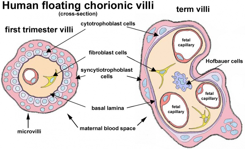
Human Floating Chorionic Villi
A figure showing the changes in placental villi between early (first trimester) and late (third trimester) placental development. Note Hofbauer cells occur in villi in both trimesters, though only shown in the later villi.
- Links: Hofbauer cells | placental villi
Reference
Figure based Fig 1.B Malassiné et al. Retrovirology 2008 5:6 doi:10.1186/1742-4690-5-6.
Cite this page: Hill, M.A. (2024, June 19) Embryology Human placental villi cartoon 01.jpg. Retrieved from https://embryology.med.unsw.edu.au/embryology/index.php/File:Human_placental_villi_cartoon_01.jpg
- © Dr Mark Hill 2024, UNSW Embryology ISBN: 978 0 7334 2609 4 - UNSW CRICOS Provider Code No. 00098G
File history
Yi efo/eka'e gwa ebo wo le nyangagi wuncin ye kamina wunga tinya nan
| Gwalagizhi | Nyangagi | Dimensions | User | Comment | |
|---|---|---|---|---|---|
| current | 12:13, 3 June 2012 | | 1,084 × 663 (142 KB) | Z8600021 (talk | contribs) | ==Human Floating Chorionic Villi== ===Reference=== Figure based Fig 1. Malassiné et al. [http://www.retrovirology.com/content/5/1/6 Retrovirology] 2008 5:6 doi:10.1186/1742-4690-5-6. |
You cannot overwrite this file.