File:Aorta coarctation MRI.jpg
Aorta_coarctation_MRI.jpg (455 × 423 pixels, file size: 25 KB, MIME type: image/jpeg)
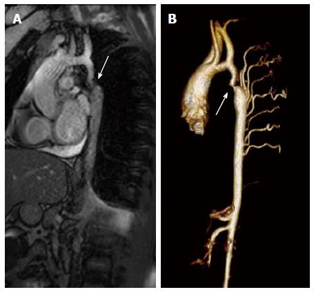
Coarctation of the Aorta Magnetic resonance imaging
A Magnetic resonance image (steady-state free precession) in a sagittal projection demonstrating transverse arch hypoplasia and long segment coarctation of the aorta distal to the left subclavian artery (arrow) in a 12-year-old male.
B Three-dimensional reconstruction of a gated contrasted angiogram for the same patient, which demonstrates transverse arch hypoplasia, coarctation at the aorta at the distal transverse aortic arch and isthmus (arrow), and dilated intercostal arteries (collaterals).
Reference
Torok RD, Campbell MJ, Fleming GA & Hill KD. (2015). Coarctation of the aorta: Management from infancy to adulthood. World J Cardiol , 7, 765-75. PMID: 26635924 DOI.
Copyright
Open-Access: This article is an open-access article which was selected by an in-house editor and fully peer-reviewed by external reviewers. It is distributed in accordance with the Creative Commons Attribution Non Commercial (CC BY-NC 4.0) license, which permits others to distribute, remix, adapt, build upon this work non-commercially, and license their derivative works on different terms, provided the original work is properly cited and the use is non-commercial. See: http://creativecommons.org/licenses/by-nc/4.0/
Figure 2 WJC-7-765-g002.jpg
Cite this page: Hill, M.A. (2024, June 26) Embryology Aorta coarctation MRI.jpg. Retrieved from https://embryology.med.unsw.edu.au/embryology/index.php/File:Aorta_coarctation_MRI.jpg
- © Dr Mark Hill 2024, UNSW Embryology ISBN: 978 0 7334 2609 4 - UNSW CRICOS Provider Code No. 00098G
File history
Yi efo/eka'e gwa ebo wo le nyangagi wuncin ye kamina wunga tinya nan
| Gwalagizhi | Nyangagi | Dimensions | User | Comment | |
|---|---|---|---|---|---|
| current | 14:51, 8 March 2019 | | 455 × 423 (25 KB) | Z8600021 (talk | contribs) | ==Coarctation of the Aorta Magnetic resonance imaging== A: Magnetic resonance image (steady-state free precession) in a sagittal projection demonstrating transverse arch hypoplasia and long segment coarctation of the aorta distal to the left subclavian... |
You cannot overwrite this file.
File usage
The following page uses this file: