File:Hassall1849 plate41 fig10.jpg
Original file (364 × 1,000 pixels, file size: 73 KB, MIME type: image/jpeg)
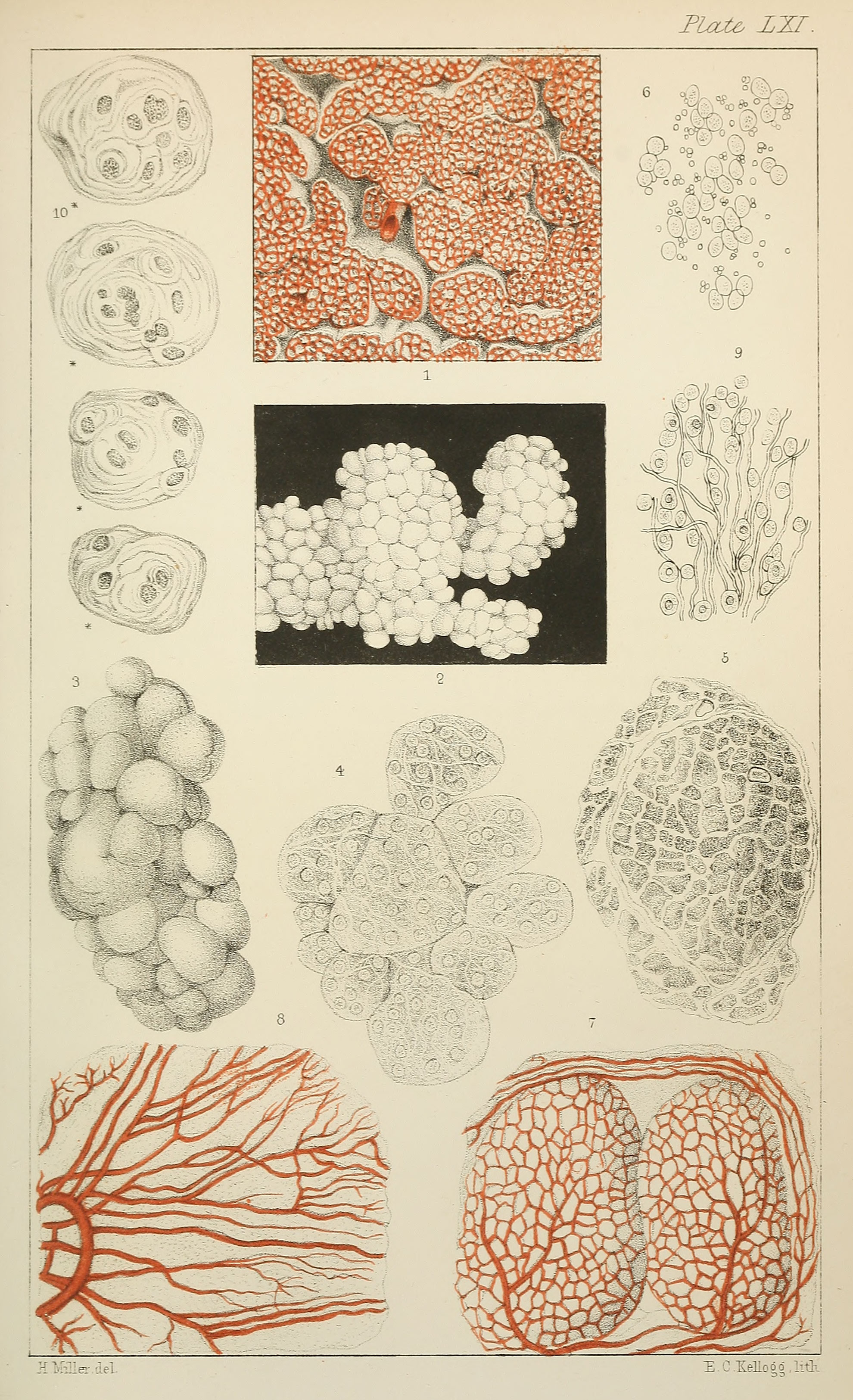
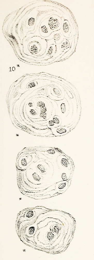
Plate LXI. Fig. 10. Compound cells of thymus
Magnified 378 diameters.
Online Editor - This historic textbook figure is often cited as the original source for "Hassall's corpuscles" within the thymus. original text source for identification of "Hassall's corpuscles" within the thymus.
Original scanned file name - Microscopicanato12hass_orig_0811.jp2
Reference
Hassall AH. The microscopic anatomy of the human body, in health and disease. (1849) Samuel Hurley, Fleet Street, London.
Cite this page: Hill, M.A. (2026, October 5) Embryology Hassall1849 plate41 fig10.jpg. Retrieved from https://embryology.med.unsw.edu.au/embryology/index.php/File:Hassall1849_plate41_fig10.jpg
- © Dr Mark Hill 2026, UNSW Embryology ISBN: 978 0 7334 2609 4 - UNSW CRICOS Provider Code No. 00098G
File history
Yi efo/eka'e gwa ebo wo le nyangagi wuncin ye kamina wunga tinya nan
| Gwalagizhi | Nyangagi | Dimensions | User | Comment | |
|---|---|---|---|---|---|
| current | 12:41, 22 Ocak 2019 | | 364 × 1,000 (73 KB) | Z8600021 (talk | contribs) | |
| 12:37, 22 Ocak 2019 | 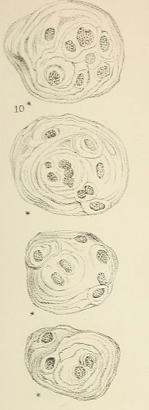 | 500 × 1,375 (101 KB) | Z8600021 (talk | contribs) |
You cannot overwrite this file.
File usage
The following 2 pages use this file: