File:HansonAnson1962 fig01.jpg
Original file (1,280 × 643 pixels, file size: 218 KB, MIME type: image/jpeg)
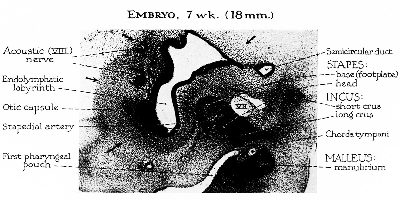
Fig. 1. The malleus Embryo 7 week 18mm
The malleus, like the incus and stapes, develops from a mesenchymal blastema located within the first two branchial arches. In the earliest stages of differentiation (approximately 10 mm) the malleus is an indistinctly outlined component of this cellular blastema.
Reference
Hanson JR. and Anson BJ. Development of the malleus of the human ear; Illustrated in atlas series. (1962) Q Bull Northwest Univ Med Sch. 36(2): 119–137. PMID: 13904457.
Cite this page: Hill, M.A. (2026, October 3) Embryology HansonAnson1962 fig01.jpg. Retrieved from https://embryology.med.unsw.edu.au/embryology/index.php/File:HansonAnson1962_fig01.jpg
- © Dr Mark Hill 2026, UNSW Embryology ISBN: 978 0 7334 2609 4 - UNSW CRICOS Provider Code No. 00098G
File history
Yi efo/eka'e gwa ebo wo le nyangagi wuncin ye kamina wunga tinya nan
| Gwalagizhi | Nyangagi | Dimensions | User | Comment | |
|---|---|---|---|---|---|
| current | 10:10, 7 Ocak 2019 | | 1,280 × 643 (218 KB) | Z8600021 (talk | contribs) | |
| 10:07, 7 Ocak 2019 | 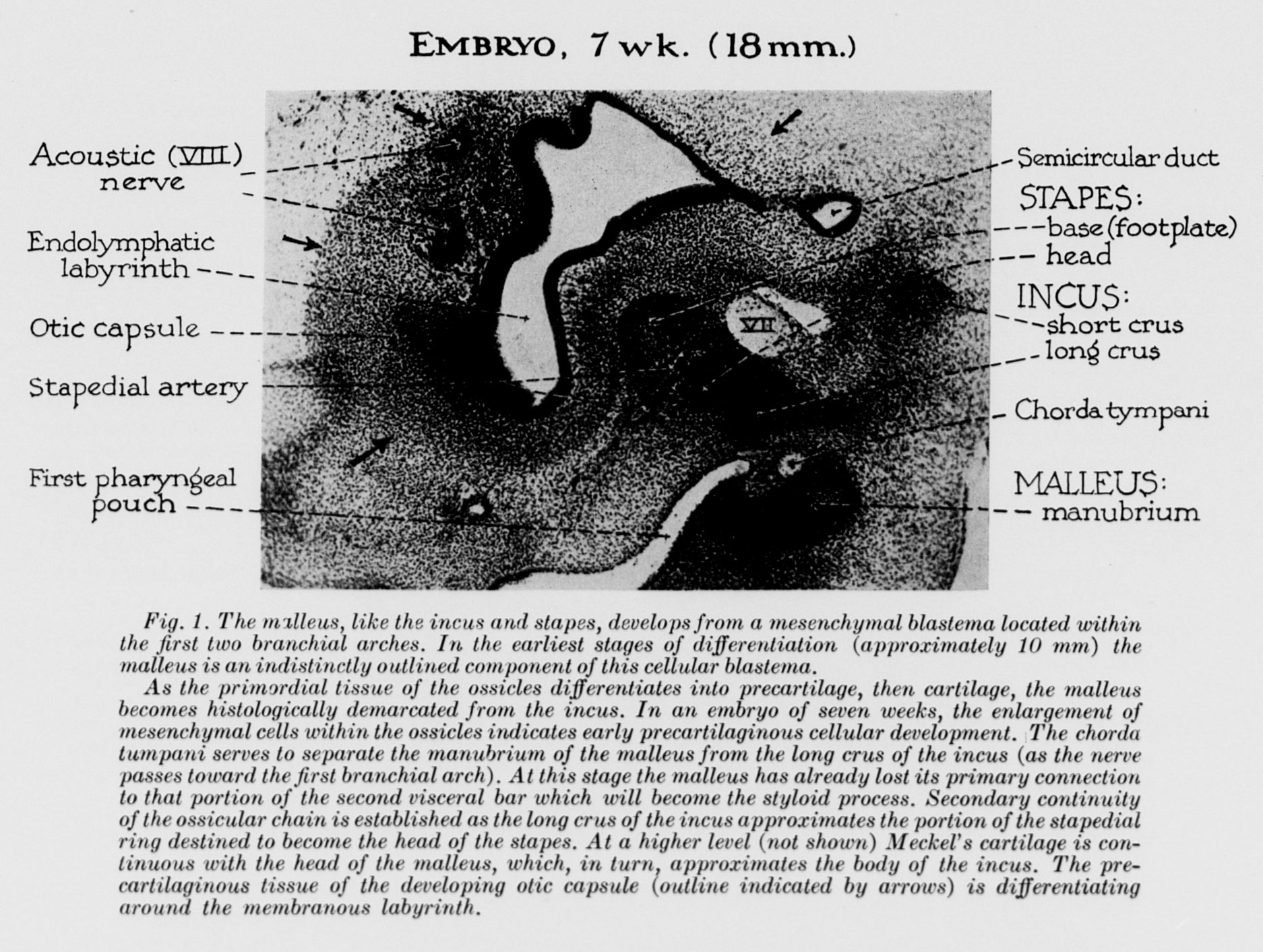 | 1,874 × 1,413 (484 KB) | Z8600021 (talk | contribs) | ===Reference=== {{Ref-HansonAnson1962}} Category:Middle Ear |
You cannot overwrite this file.
File usage
The following 2 pages use this file:


