File:Shanklin1940 fig02.jpg
From Embryology
Size of this preview: 800 × 546 pixels. Other resolution: 1,000 × 682 pixels.
Original file (1,000 × 682 pixels, file size: 131 KB, MIME type: image/jpeg)
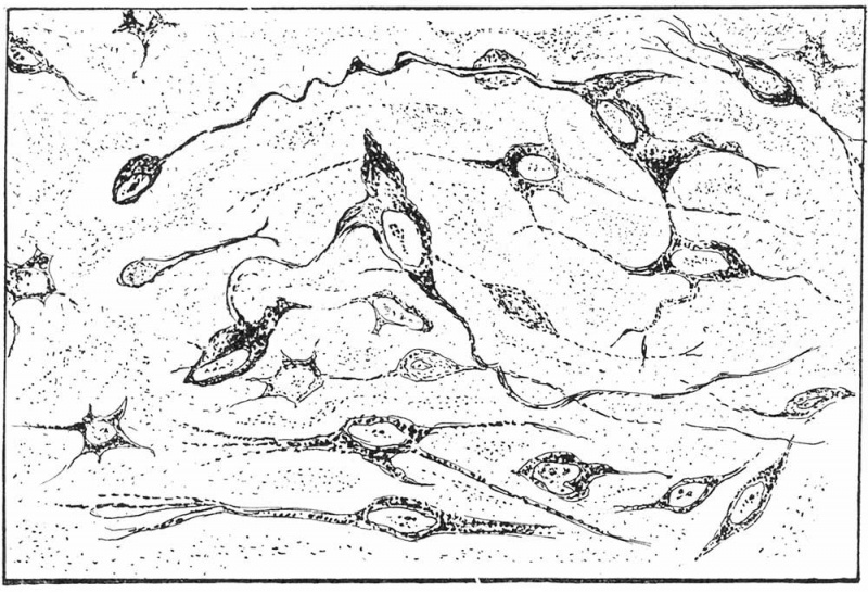
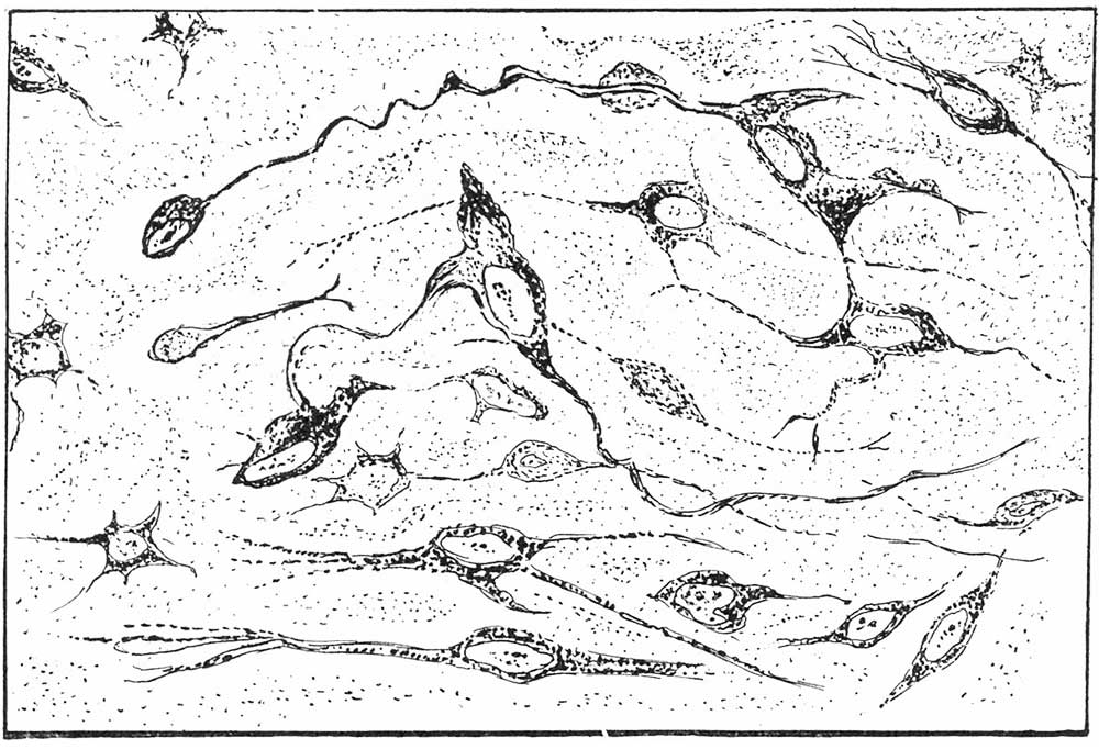
Text-fig. 2. Drawing of pituicytes of a 7.5 month human
Premature new-born that lived for 2 months. Pituicytes show anastomosis between processes, granules along one of the processes, also a bulb-like ending on one of the terminals.
Reference
Shanklin WM. Differentiation of pituicytes in the human foetus. (1940) J Anat. 74(4): 459-63. PMID 17104829
Cite this page: Hill, M.A. (2026, October 3) Embryology Shanklin1940 fig02.jpg. Retrieved from https://embryology.med.unsw.edu.au/embryology/index.php/File:Shanklin1940_fig02.jpg
- © Dr Mark Hill 2026, UNSW Embryology ISBN: 978 0 7334 2609 4 - UNSW CRICOS Provider Code No. 00098G
File history
Yi efo/eka'e gwa ebo wo le nyangagi wuncin ye kamina wunga tinya nan
| Gwalagizhi | Nyangagi | Dimensions | User | Comment | |
|---|---|---|---|---|---|
| current | 22:28, 26 Haziran 2018 | | 1,000 × 682 (131 KB) | Z8600021 (talk | contribs) | |
| 22:27, 26 Haziran 2018 | 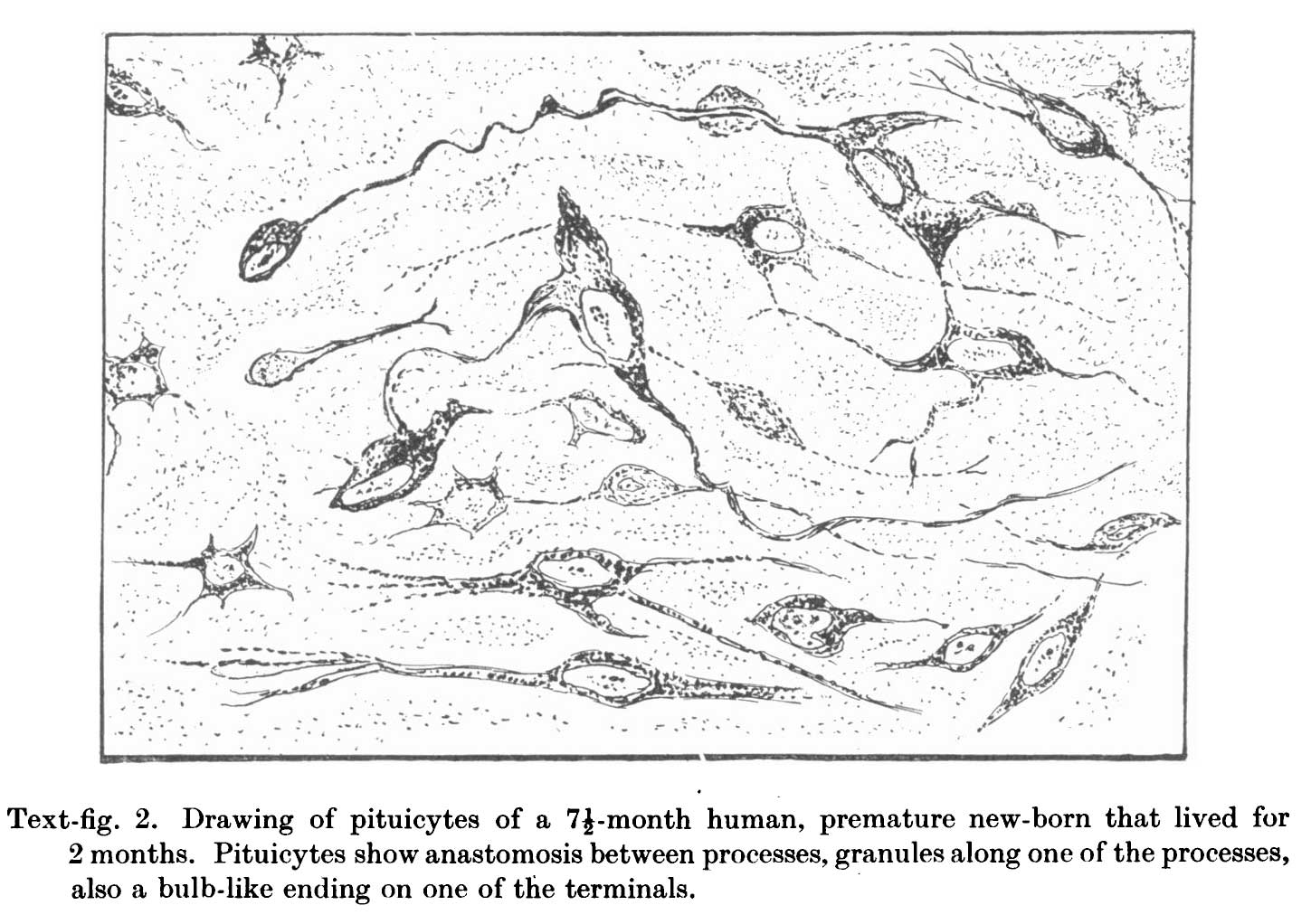 | 1,449 × 1,022 (167 KB) | Z8600021 (talk | contribs) |
You cannot overwrite this file.
File usage
The following page uses this file: