File:Frazer1919 fig02.jpg
From Embryology
Size of this preview: 800 × 514 pixels. Other resolution: 1,280 × 823 pixels.
Original file (1,280 × 823 pixels, file size: 144 KB, MIME type: image/jpeg)
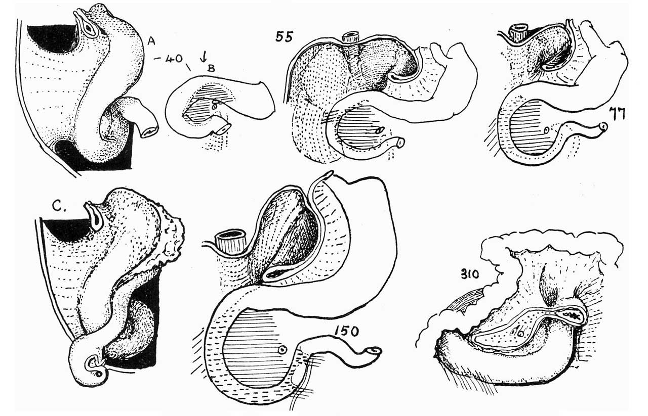
Fig. 2. Side and front views of 40mm model. Front views of older specimens
0, a semidiagrammatic drawing to show position of colon at end of second stage. In the later stages shown, pancreas is indicated by horizontal shading, adhesions by oblique shading and interrupted lines.
File history
Yi efo/eka'e gwa ebo wo le nyangagi wuncin ye kamina wunga tinya nan
| Gwalagizhi | Nyangagi | Dimensions | User | Comment | |
|---|---|---|---|---|---|
| current | 14:12, 15 Haziran 2018 | | 1,280 × 823 (144 KB) | Z8600021 (talk | contribs) | |
| 14:10, 15 Haziran 2018 | 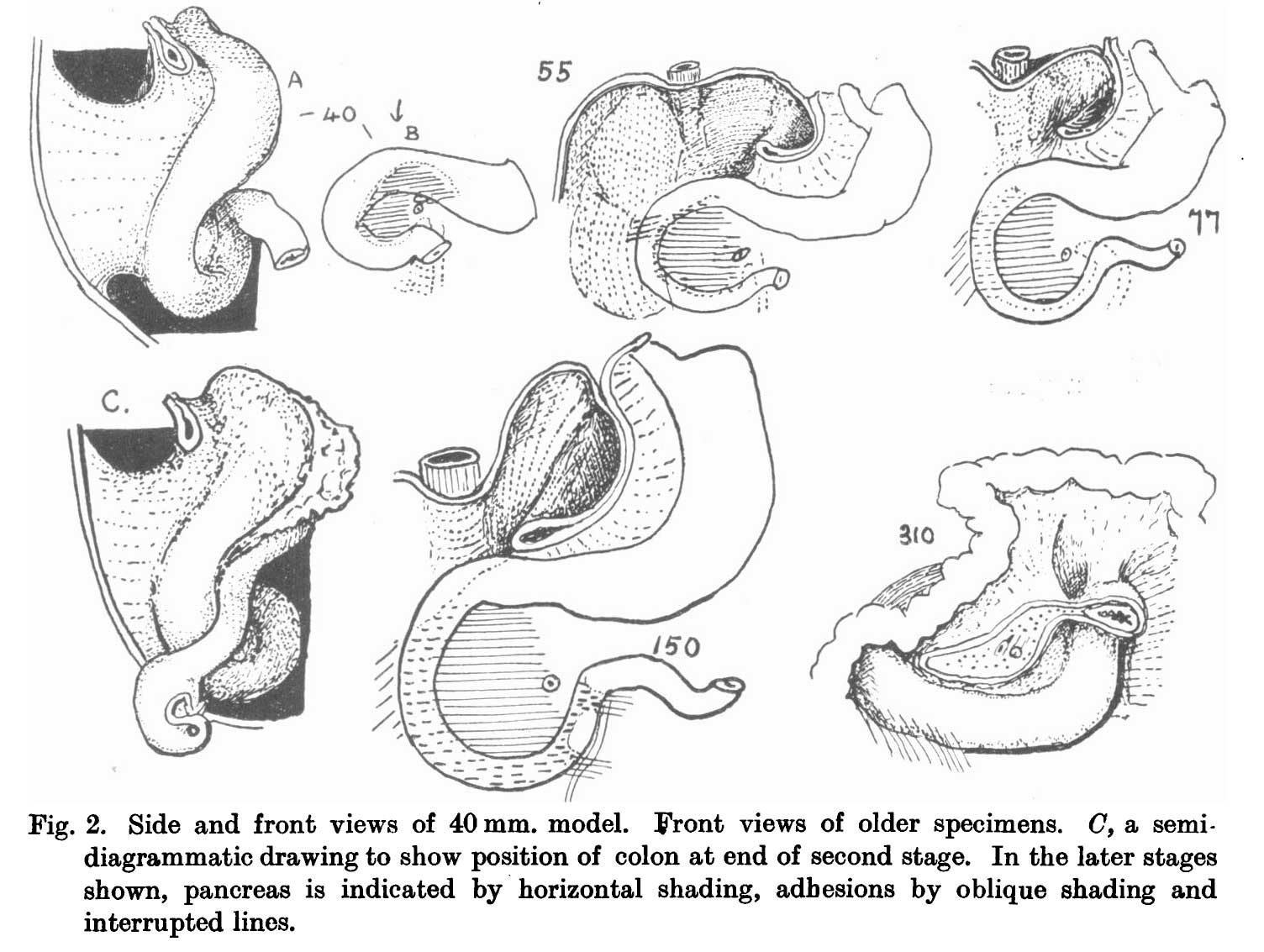 | 1,504 × 1,133 (174 KB) | Z8600021 (talk | contribs) |
You cannot overwrite this file.
File usage
The following page uses this file:


