File:Cooper1932-fig03.jpg
From Embryology
Size of this preview: 620 × 599 pixels. Other resolution: 901 × 871 pixels.
Original file (901 × 871 pixels, file size: 211 KB, MIME type: image/jpeg)
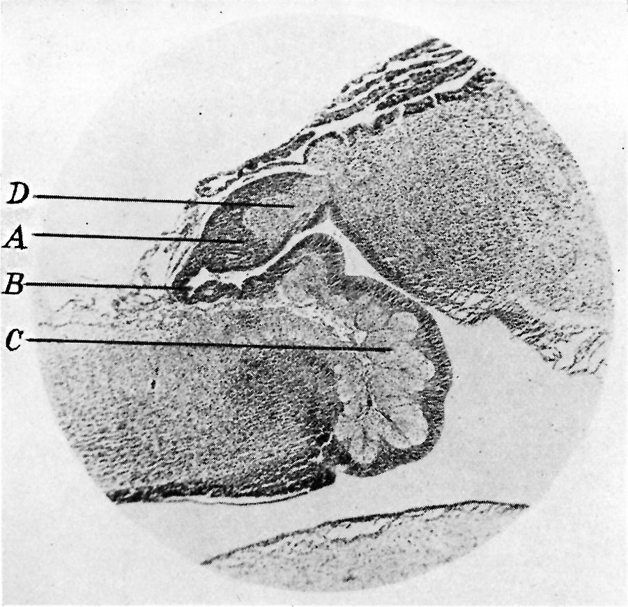
Fig. 3. Sagittal section through pineal region in embryo between the third and fourth months
x 35. A, anterior anlage of pineal. B, posterior anlage with pineel diverticulum. C, posterior commissure. D, habenular commissure.
| Historic Disclaimer - information about historic embryology pages |
|---|
 |
- Links: pineal
Reference
Cooper ERA. The human pineal gland and pineal cysts. (1932)
Cite this page: Hill, M.A. (2024, June 27) Embryology Cooper1932-fig03.jpg. Retrieved from https://embryology.med.unsw.edu.au/embryology/index.php/File:Cooper1932-fig03.jpg
- © Dr Mark Hill 2024, UNSW Embryology ISBN: 978 0 7334 2609 4 - UNSW CRICOS Provider Code No. 00098G
File history
Yi efo/eka'e gwa ebo wo le nyangagi wuncin ye kamina wunga tinya nan
| Gwalagizhi | Nyangagi | Dimensions | User | Comment | |
|---|---|---|---|---|---|
| current | 17:26, 21 May 2018 | | 901 × 871 (211 KB) | Z8600021 (talk | contribs) | |
| 17:24, 21 May 2018 |  | 1,470 × 1,077 (298 KB) | Z8600021 (talk | contribs) |
You cannot overwrite this file.
File usage
The following 2 pages use this file: