File:Gilmour1941 plate10.jpg
Original file (1,501 × 2,265 pixels, file size: 391 KB, MIME type: image/jpeg)
Plate X
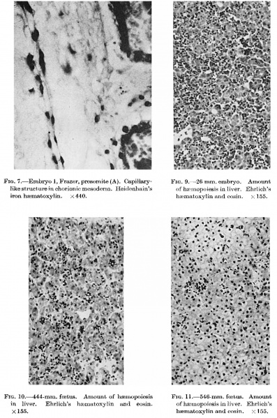
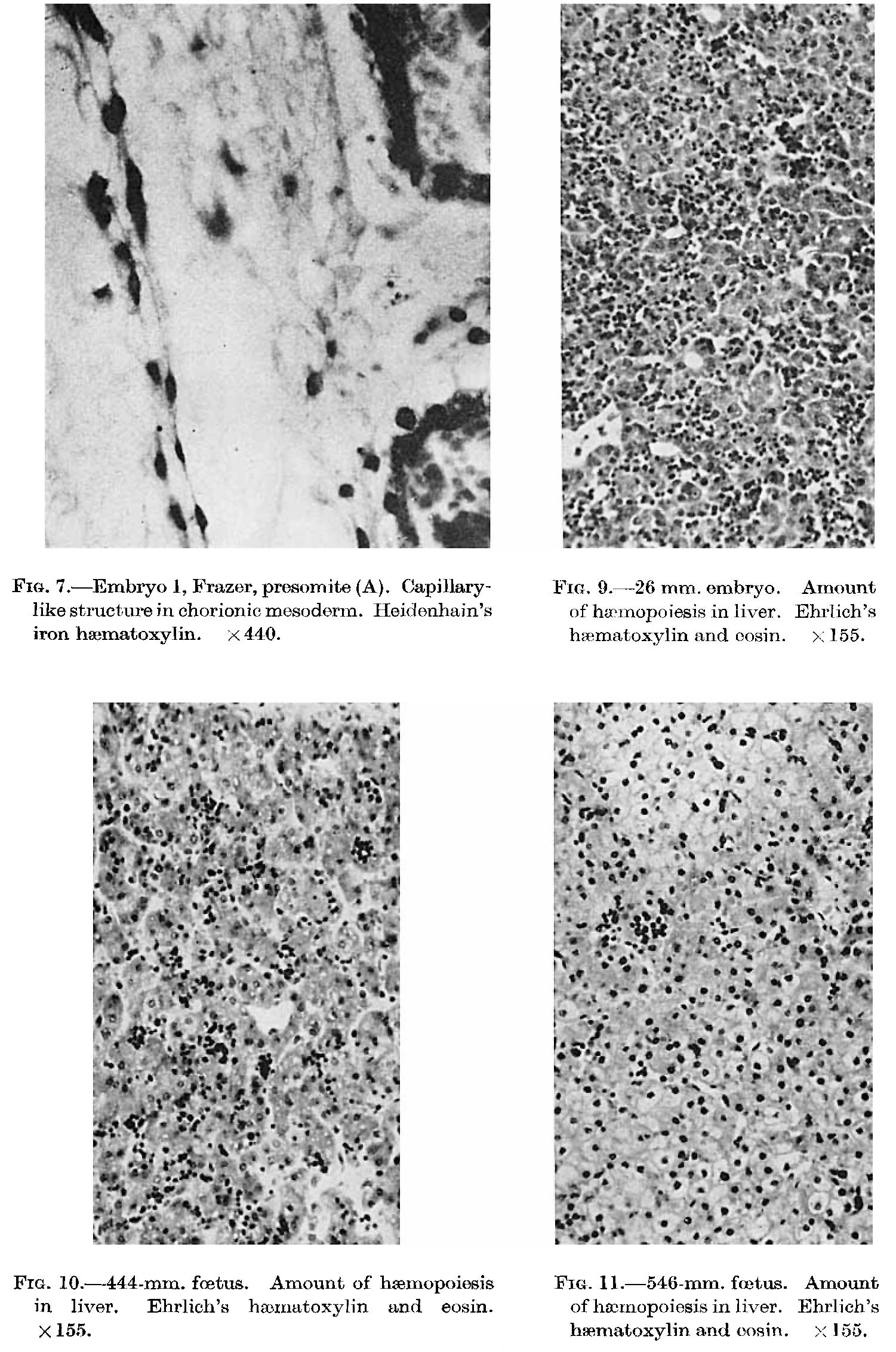
Fig. 7. Embryo 1, Frazer, presornite (A). Capi11ary
Fig. 9. 26 mm. embryo. Amount like structure in chorionie mesoderm HeideI1hai11’s of hzmnopoiesis in liver. Ehr1ich’s haematoxylin and eosin. X 155.
iron haematoxylin. X 440.
FIG. 10. 444-mm. foetus. Amount of haemopoiesis
FIG. 1l.—546—mm. fmt-us. Amount Ehrlich’s hae.xI1a.t0xy11'n and eosin.
of hwmopoiesis in liver. Ehrlich’s
in liver. X 155.
haematoxylin. and eosin.
x I55.
Reference
Gilmour JR. Normal haemopoiesis in intra-uterine and neonatal life. (1941) J. Pathol. Bacteriol. 52: 25-55.
Cite this page: Hill, M.A. (2026, October 7) Embryology Gilmour1941 plate10.jpg. Retrieved from https://embryology.med.unsw.edu.au/embryology/index.php/File:Gilmour1941_plate10.jpg
- © Dr Mark Hill 2026, UNSW Embryology ISBN: 978 0 7334 2609 4 - UNSW CRICOS Provider Code No. 00098G
File history
Yi efo/eka'e gwa ebo wo le nyangagi wuncin ye kamina wunga tinya nan
| Gwalagizhi | Nyangagi | Dimensions | User | Comment | |
|---|---|---|---|---|---|
| current | 11:29, 17 Mayıs 2018 | | 1,501 × 2,265 (391 KB) | Z8600021 (talk | contribs) | |
| 11:24, 17 Mayıs 2018 | 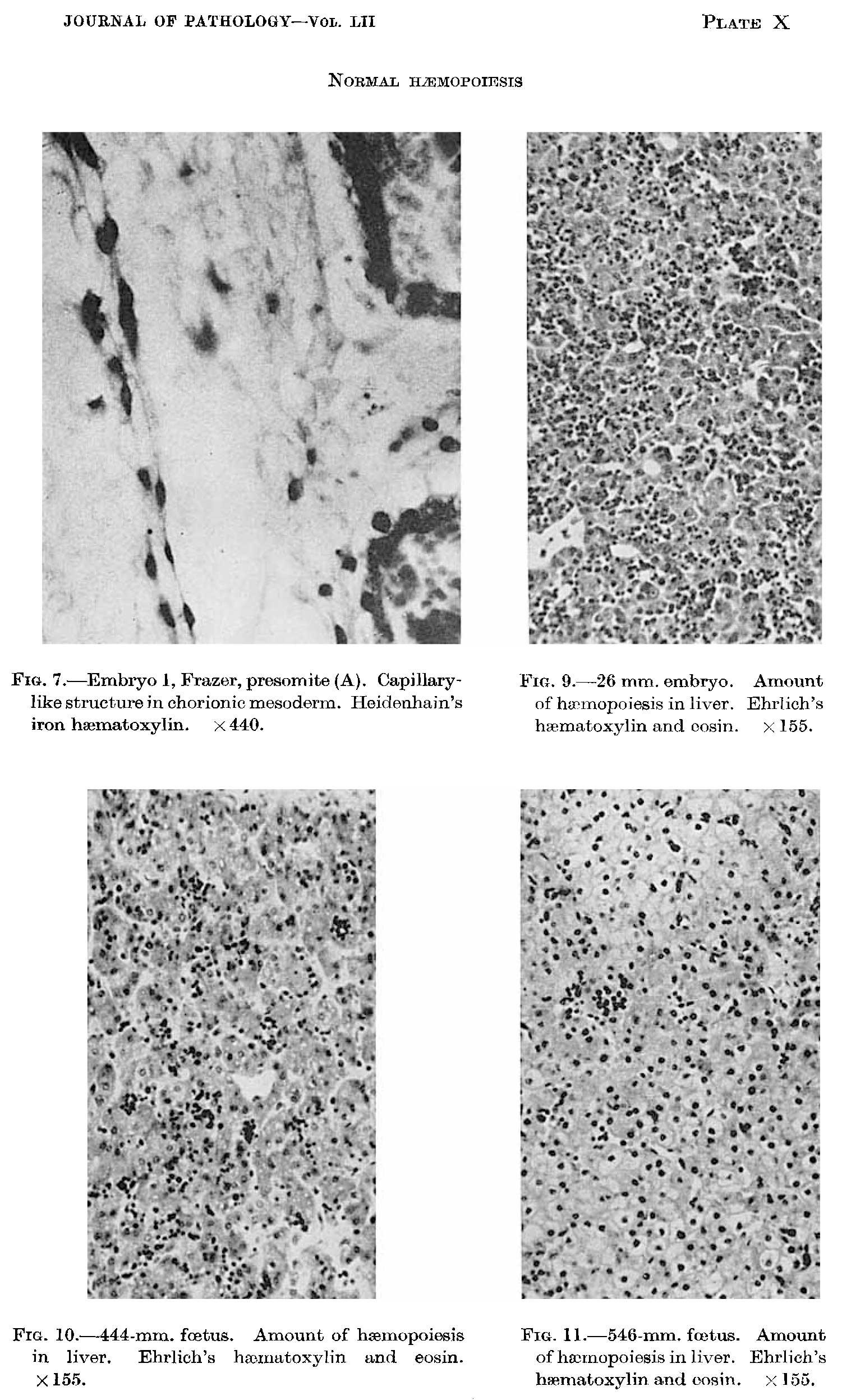 | 1,501 × 2,494 (398 KB) | Z8600021 (talk | contribs) |
You cannot overwrite this file.
File usage
The following page uses this file: