File:Anderson2016-fig43a.jpg
From Embryology

Size of this preview: 600 × 600 pixels.
Original file (800 × 800 pixels, file size: 60 KB, MIME type: image/jpeg)
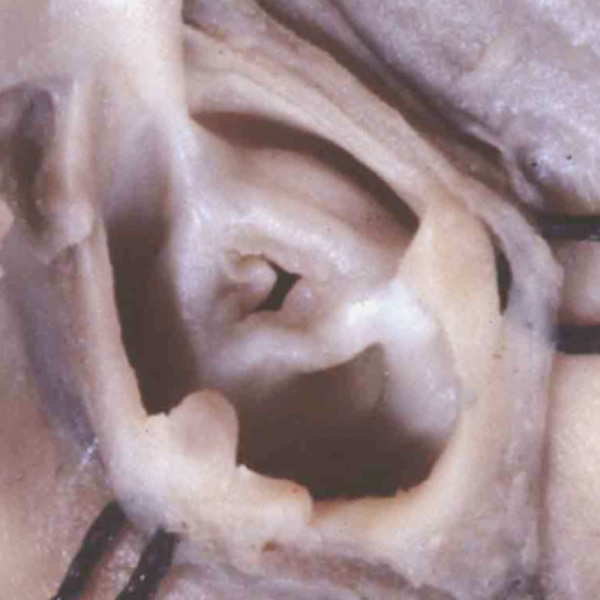
Fig. 43a. Stenotic Pulmonary Valve
The images show, to the left hand, a critically stenotic pulmonary valve, and to the right hand, a critically stenotic aortic valve. These lesions are well explained on the basis of excessive fusion of the distal outflow cushions and the intercalated cushion during the fifth or sixth week of development, or at any stage onwards to term. It is likely that the changes to produce the stenotic valves occur later in development, but the initial insult could occur during the fifth or sixth week.
File history
Click on a date/time to view the file as it appeared at that time.
| Date/Time | Thumbnail | Dimensions | User | Comment | |
|---|---|---|---|---|---|
| current | 23:01, 16 February 2017 | | 800 × 800 (60 KB) | Z8600021 (talk | contribs) |
You cannot overwrite this file.