File:Keibel Mall 2 629.jpg
From Embryology
Size of this preview: 800 × 501 pixels. Other resolution: 1,000 × 626 pixels.
Original file (1,000 × 626 pixels, file size: 118 KB, MIME type: image/jpeg)
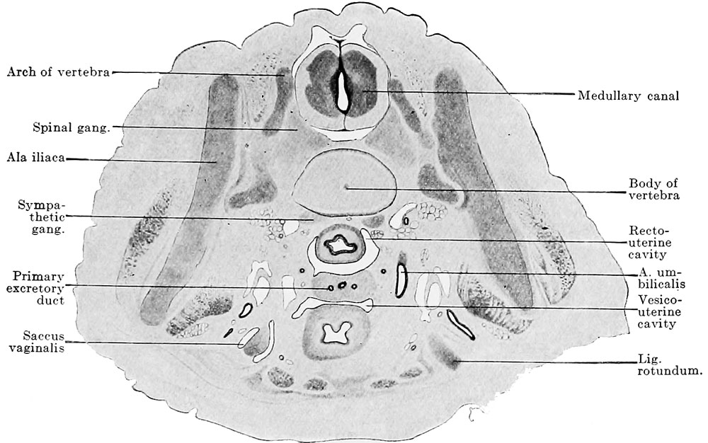
Fig. 629. Transverse section of a human embryo of 32 mm vertex-breech length at the level of the utero-vaginal canal
(Embryo 426, I, IS in the collection of the Anatomical Institute, Zurich, IX. 1, 2.) X 15.
The section shows the genital cord stretched transversely through the cavity of the true pelvis, and in it are the two primary excretory ducts and the utero-vaginal canal. The mesenchymatous uterine wall, which encloses the two primary excretory ducts, is so situated that it divides the genital cord into three portions, the uterus and the two lig. lata.
File history
Yi efo/eka'e gwa ebo wo le nyangagi wuncin ye kamina wunga tinya nan
| Gwalagizhi | Nyangagi | Dimensions | User | Comment | |
|---|---|---|---|---|---|
| current | 22:29, 14 February 2017 | | 1,000 × 626 (118 KB) | Z8600021 (talk | contribs) | |
| 22:28, 14 February 2017 | 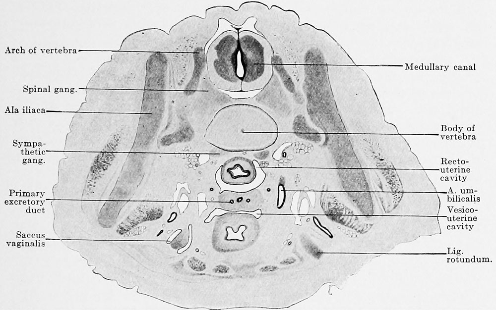 | 1,000 × 626 (122 KB) | Z8600021 (talk | contribs) | ||
| 22:24, 14 February 2017 | 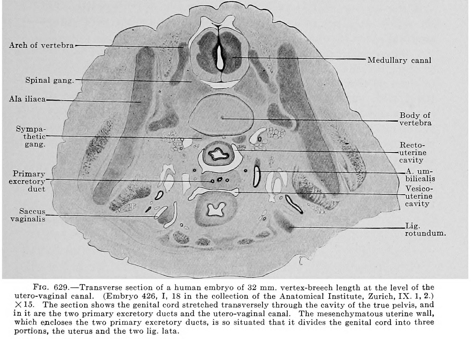 | 1,564 × 1,124 (348 KB) | Z8600021 (talk | contribs) |
You cannot overwrite this file.
File usage
The following 2 pages use this file: