File:Kramer1942 fig01.jpg
Original file (1,280 × 1,108 pixels, file size: 280 KB, MIME type: image/jpeg)
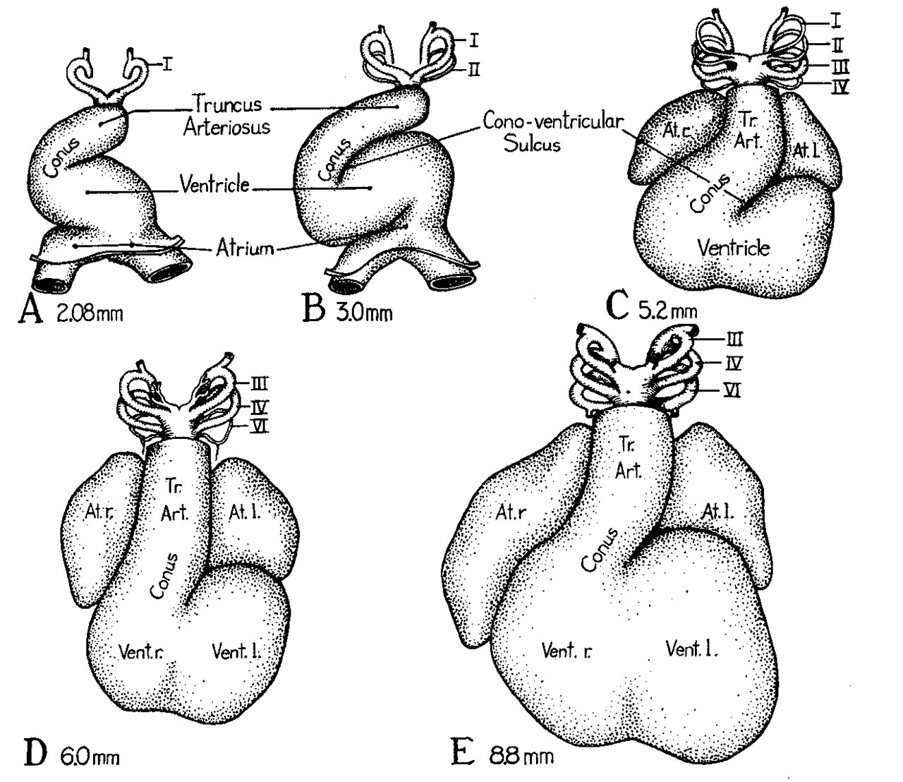
Fig. 1. Ventral views of reconstructions of the hearts of young embryos
Showing the shift in the position of the truncus toward the mid-line and the reduction of the cono-ventricular sulcns. Slight modifications in the redrawing have been made in order to bring the various models into comparable orientation.
A 2.08 mm. embryo X 42 (Modified from Davis, '27, fig. 23).
B 3 mm. embryo X 42 (Modified from Tandler, ’12, fig. 374).
C 5.2 mm. embryo X 42 (Modified from Tandler, '12, fig. 378).
D 6.0 mm. embryo X 21 (Modified from Waterston, ’18, fig. 2).
E 8.8 mm. embryo X21 (Original figure from reconstruction of EH. 35, University of Michigan Coll.).
Reference
Kramer TC. The partitioning of the truncus and conus and the formation of the membranous portion of the interventricular septum in the human heart. (1942) Amer. J Anat. 71(3): 343-370.
File history
Yi efo/eka'e gwa ebo wo le nyangagi wuncin ye kamina wunga tinya nan
| Gwalagizhi | Nyangagi | Dimensions | User | Comment | |
|---|---|---|---|---|---|
| current | 11:46, 4 February 2017 | | 1,280 × 1,108 (280 KB) | Z8600021 (talk | contribs) | |
| 09:15, 4 February 2017 | 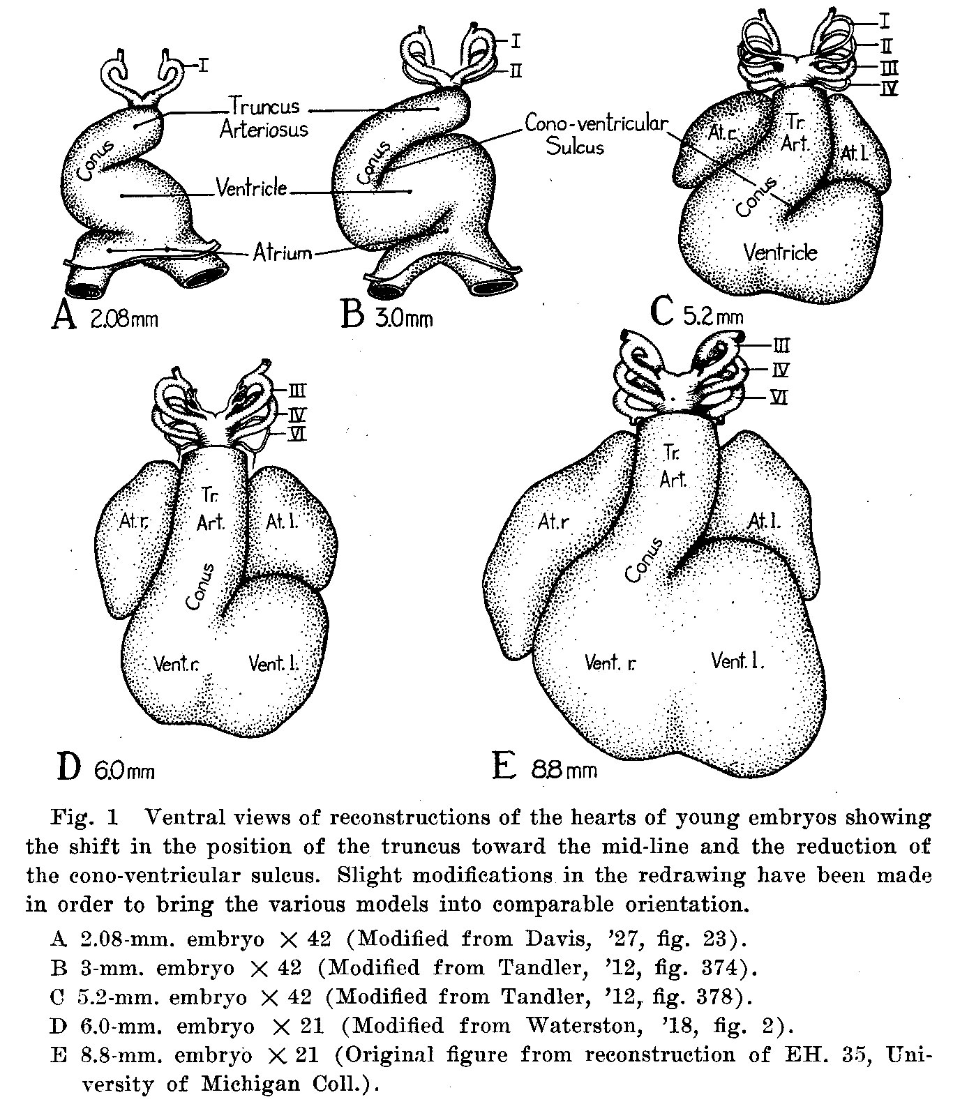 | 1,366 × 1,593 (477 KB) | Z8600021 (talk | contribs) | ===Reference=== {{Ref-Kramer1942}} |
You cannot overwrite this file.
File usage
The following page uses this file:


