File:Hunter1935 text-fig01.jpg
From Embryology
Size of this preview: 478 × 599 pixels. Other resolution: 488 × 612 pixels.
Original file (488 × 612 pixels, file size: 31 KB, MIME type: image/jpeg)
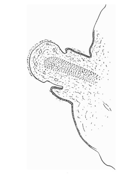
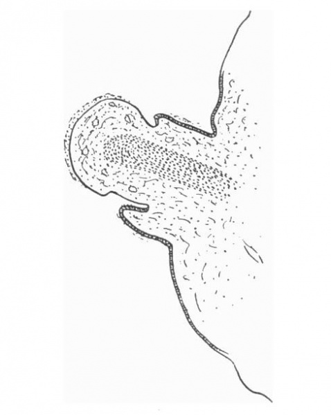
Text-fig. 1. L.S. Penis of human foetus 40 mm C.R. length
Note the ridge of ectoderm behind the corona and the glans, and the thin layer of desquamating cells on the surface.
Reference
Hunter RH. Notes on the development of the prepuce. (1935) J Anat. 70: 68-75. PMID 17104576
Cite this page: Hill, M.A. (2024, June 22) Embryology Hunter1935 text-fig01.jpg. Retrieved from https://embryology.med.unsw.edu.au/embryology/index.php/File:Hunter1935_text-fig01.jpg
- © Dr Mark Hill 2024, UNSW Embryology ISBN: 978 0 7334 2609 4 - UNSW CRICOS Provider Code No. 00098G
File history
Yi efo/eka'e gwa ebo wo le nyangagi wuncin ye kamina wunga tinya nan
| Gwalagizhi | Nyangagi | Dimensions | User | Comment | |
|---|---|---|---|---|---|
| current | 12:29, 4 January 2017 | | 488 × 612 (31 KB) | Z8600021 (talk | contribs) | |
| 12:29, 4 January 2017 | 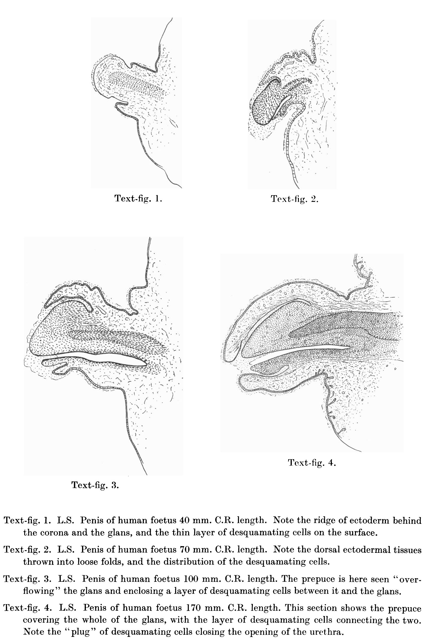 | 1,454 × 2,174 (438 KB) | Z8600021 (talk | contribs) | ===Reference=== {{Ref-Hunter1935}} |
You cannot overwrite this file.
File usage
The following 2 pages use this file: