File:Rat- pancreatic islet development.jpg

Original file (1,200 × 524 pixels, file size: 230 KB, MIME type: image/jpeg)
Rat- pancreatic islet development
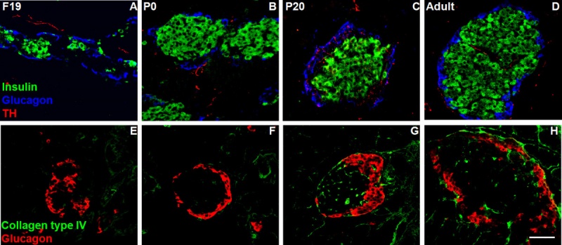
Morphological reorganization of pancreatic islets from prenatal life to adulthood.
We localised sympathetic nerve fibres and collagen capsules surrounding the islets by confocal microscopy in longitudinal sections of pancreases.
Immunostaining for insulin (green), glucagon (blue), tyrosine hydroxylase (TH; red) and collagen type IV (2nd line, green) at different developmental rat stages (F19, P1, P20 and adult). Scale bar = 50 μm.
1471-213X-9-34-1-l.jpg
Reference
<pubmed>19534767</pubmed>
Cabrera-Vásquez et al. BMC Developmental Biology 2009 9:34 doi:10.1186/1471-213X-9-34
© 2009 Cabrera-Vásquez et al; licensee BioMed Central Ltd. This is an Open Access article distributed under the terms of the Creative Commons Attribution License (http://creativecommons.org/licenses/by/2.0), which permits unrestricted use, distribution, and reproduction in any medium, provided the original work is properly cited.
File history
Yi efo/eka'e gwa ebo wo le nyangagi wuncin ye kamina wunga tinya nan
| Gwalagizhi | Nyangagi | Dimensions | User | Comment | |
|---|---|---|---|---|---|
| current | 16:09, 18 Mayıs 2010 | | 1,200 × 524 (230 KB) | S8600021 (talk | contribs) | ==Rat- pancreatic islet development== Morphological reorganization of pancreatic islets from prenatal life to adulthood. We localised sympathetic nerve fibres and collagen capsules surrounding the islets by confocal microscopy in longitudinal sections |
You cannot overwrite this file.
File usage
The following page uses this file: