File:Reagan1912 fig09-15.jpg
Original file (1,581 × 1,895 pixels, file size: 222 KB, MIME type: image/jpeg)
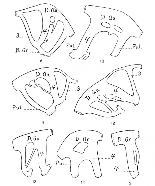
Fig. 9 to 15.
Fig. 9 The aortic arches of t he left side of an 8.5 mm. pig. (The development of the rudimentary vessel seems to have been impeded by the common base of the glandulethyroidicnne and the pre-pulmonic caecum).
Fig. 10 The left fourth and pulmonic arches of the S.6 mm. pig. No. 78.S of the C. H. Spurgeon collection. (No attempt was made to illustrate these conditions in figs. 6 and 7.)
Fig. 11 Aortic arches of the right side of a 9.5 mm. pig, showing irregular conditions.
Fig. 12 Aortic arches of the right side of a 10 nun. pig, showing unusual conditions.
Fig. 13 The fourth and pulmonic arches of the right side of a 10 mm. pig, showing the connection between the base of the pulmonic and the dorsal part of the fourth. (Conditions similar to this were observed in three instances).
Fig. 14. The fourth and pulmonic arches of a 9. (> nun. pig, showing a broad connection between the dorsal portions.
Fig. 15 The fourth arch of a 6.5 mm. pig, showing a division of that vessel on its anterior face.
File history
Yi efo/eka'e gwa ebo wo le nyangagi wuncin ye kamina wunga tinya nan
| Gwalagizhi | Nyangagi | Dimensions | User | Comment | |
|---|---|---|---|---|---|
| current | 14:19, 13 Haziran 2016 | | 1,581 × 1,895 (222 KB) | Z8600021 (talk | contribs) |
You cannot overwrite this file.
File usage
The following page uses this file: