Talk:Draft 2016

From Embryology
Revision as of 15:59, 26 February 2016 by Z8600021 (talk | contribs)

Spleen

Thymus Anatomy

Thymus Anatomy
Thymus cartoon.jpg Gray1178.jpg
adult thymus - bilobed, superior mediastinum, anterior to heart infant thymus - large
Thymus Involution
Fetal thymus weight growth graph.jpg Overall Size Changes with age
  • birth 10-15 g
  • puberty 30-40 g
  • after puberty - involution
    • replaced by adipose tissue
  • middle-aged 10 g

Thymus Functions

Thymus Functions 
  • specialised thymus microenvironments allow the production of self-tolerant T-cells from immature precursors.
  • release into circulation of these cells
  • destruction of cells that recognise self antigens

Thymus Structure

Thymus Structure 
Spleen structure cartoon 01.jpg


Thymus Histology

Thymus Histology 
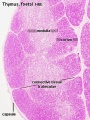
  • Capsule with trabeculae (dense connective tissue)

Spleen histology 06.jpg


Overview - red and white pulp Overview - blood vessels Red pulp
Spleen histology 01.jpg Spleen histology 02.jpg Spleen histology 03.jpg

Reticular Fibers (type III collagen) act as supporting meshwork (can be seen in Silver stained preparations)

Spleen histology 05.jpg Spleen histology 04.jpg
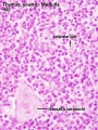
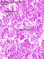
Thymus Histology: Fetal Thymus overview | Fetal Thymus Medulla | Fetal Thymus Cortex | Adult Thymus | unlabeled fetal overview | unlabeled fetal medulla |unlabeled fetal thymic corpuscle |unlabeled fetal cortex | unlabeled adult overview | Category:Thymus | Immune System Development


Thymus

Adult Thymus
Fetal thymus anatomy

Gross Anatomy

  • Superior mediastinum, anterior to heart
  • Bilobed lymphoepithelial organ
Histology
  • Contains reticular cells but no fibers
  • Stem lymphocytes
    • proliferate and differentiate
    • forms long-lived T- lymphocytes
  • removes auto-reactive T-lymphocytes (negative selection)
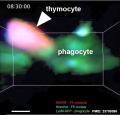
Mouse adult thymus 11.jpg
 ‎‎Thymus 1
Page | Play

Thymus Cells

Reticular cells Macrophages Lymphocytes
  • Abundant, eosinophilic, large, ovoid and light nucleus 1-2 nucleoli
  • sheathe cortical capillaries
  • form an epitheloid layer
  • maintain microenvironment for development of T-lymphocytes in cortex (thymic epitheliocytes)
  • cortex and medulla
  • difficult to distinguish from reticular cells in (Stain - Haematoxylin Eosin)
  • remove auto-reactive T-lymphocytes
  • located in cortex and medulla
  • more numerous (denser) in cortex
  • majority are developing T-lymphocytes (= thymic lymphocytes or thymocytes)


Histology

Adult Thymus

Thymus adult.jpg
  • Cortical lymphoid tissue is replaced by adipose tissue (involution)
  • Increase in size of thymic corpuscles
  • Thymic corpuscle - (Hassall’s corpuscle) mass of concentric epithelioreticular cells.

Thymus - young 01.jpg