Category:Axial Skeleton
From Embryology

This Embryology category shows media and pages related to the development of the axial skeleton (skull, vertebra, intervertebral disc and ribs).
Subcategories
This category has the following 4 subcategories, out of 4 total.
Pages in category 'Axial Skeleton'
The following 51 pages are in this category, out of 51 total.
B
P
- Paper - Development of the thoracic vertebrae in man (1905)
- Paper - Early development of the cervical vertebrae and the base of the occipital bone in man
- Paper - Embryological stages in the development of spina bifida and myeloschisis
- Paper - Fusion of cervical vertebrae (1932)
- Paper - Studies of the development of the human skeleton (1905)
- Paper - The development and reduction of the tail and of the caudal end of the spinal cord (1920)
- Paper - The sternum - its early development and ossification in man and mammals
- Paper - Vertebral Regional Determination in Young Human Embryos
- Template:Paraxial mesoderm
- Template:Primitive segment
- Template:Primitive segments
R
- Template:Ref-Bardeen1904
- Template:Ref-Bardeen1905a
- Template:Ref-Bardeen1908
- Template:Ref-Bardeen1908a
- Template:Ref-Brash1915
- Template:Ref-Fell1939
- Template:Ref-Geddes1912a
- Template:Ref-Kunitomo1920
- Template:Ref-O'Rahilly1980
- Template:Ref-O'Rahilly1983
- Template:Ref-Pan1932
- Template:Ref-Paterson1900
- Template:Ref-Paterson1902
- Template:Ref-Patten 1946b
- Template:Ref-Patten1946
- Template:Ref-Patten1953b
- Template:Ref-PMID3535481
- Template:Ref-Ruth1932
- Template:Ref-Sensenig1949
- Template:Ref-Shore1930
- Template:Ref-Wakeley1923a
- Template:Rib
- Template:Ribs
S
Media in category 'Axial Skeleton'
The following 84 files are in this category, out of 84 total.
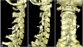 Absent cervical spine pedicle.jpg 600 × 338; 30 KB
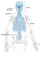 Axial skeleton.jpg 1,000 × 1,434; 111 KB
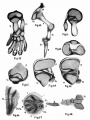 Bardeen1905 plate13.jpg 1,000 × 1,337; 85 KB
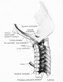 Bardeen1908a fig02.jpg 1,000 × 1,295; 85 KB
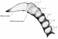 Bardeen1908a fig03.jpg 1,280 × 858; 57 KB
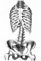 Braune 1877 plate 30 fig1.jpg 805 × 1,200; 205 KB
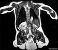 Caudal duplication syndrome.jpg 700 × 599; 47 KB
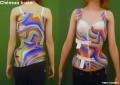 Cheneau brace.jpg 800 × 565; 79 KB
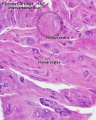 Fibrous cartilage 02.jpg 500 × 626; 96 KB
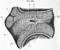 Foster135.jpg 965 × 815; 230 KB
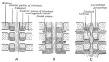 Gray0065.jpg 600 × 345; 39 KB
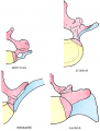 Gray0067.png 350 × 463; 46 KB
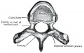 Gray0082.jpg 450 × 287; 30 KB
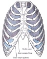 Gray0965.jpg 600 × 750; 130 KB
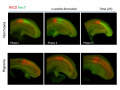 Hongjun Shi Research photo02.jpg 899 × 677; 58 KB
 Hongjun Shi Research photo03.jpg 457 × 836; 54 KB
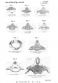 Human axial skeleton- axis development 01.jpg 763 × 1,095; 95 KB
 Human week 10 fetus 09.jpg 1,200 × 900; 345 KB
 Human week 10 fetus 11.jpg 1,200 × 900; 304 KB
 Keibel Mall 231-232.jpg 627 × 919; 70 KB
 Keibel Mall 231.jpg 450 × 511; 21 KB
 Keibel Mall 232.jpg 450 × 511; 35 KB
 Keibel Mall 260-263.jpg 726 × 800; 90 KB
 Keibel Mall 260.jpg 506 × 508; 25 KB
 Keibel Mall 261.jpg 506 × 508; 32 KB
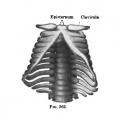 Keibel Mall 262.jpg 506 × 508; 24 KB
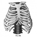 Keibel Mall 263.jpg 506 × 508; 37 KB
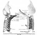 Keibel Mall 264-265.jpg 831 × 800; 88 KB
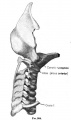 Keibel Mall 264.jpg 475 × 800; 38 KB
 Keibel Mall 265.jpg 526 × 800; 51 KB
 Keibel Mall 266.jpg 740 × 608; 77 KB
 Keibel Mall 267.jpg 500 × 410; 28 KB
 Keibel Mall 271.jpg 800 × 419; 60 KB
 Keibel Mall 273-no-legend.jpg 791 × 744; 164 KB
 Keibel Mall 273.jpg 900 × 861; 200 KB
 Keibel Mall 274-278.jpg 717 × 1,072; 123 KB
 Keith1902 fig113.jpg 675 × 1,000; 72 KB
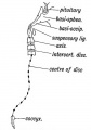 Keith1902 fig117.jpg 561 × 800; 46 KB
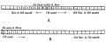 Keith1902 fig120.jpg 1,000 × 396; 54 KB
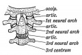 Keith1902 fig122.jpg 600 × 404; 54 KB
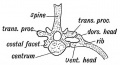 Keith1902 fig123.jpg 650 × 353; 43 KB
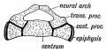 Keith1902 fig124.jpg 600 × 297; 41 KB
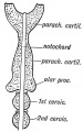 Keith1902 fig133.jpg 437 × 700; 56 KB
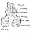 Keith1902 fig134.jpg 590 × 697; 66 KB
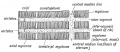 Keith1902 fig229.jpg 1,000 × 434; 106 KB
 Keith1921 fig047.jpg 537 × 1,116; 82 KB
 Keith1921 fig048.jpg 1,200 × 711; 96 KB
 Keith1921 fig049.jpg 913 × 630; 128 KB
 Keith1921 fig050.jpg 1,200 × 479; 75 KB
 Keith1921 fig051.jpg 848 × 728; 102 KB
 Keith1921 fig052.jpg 698 × 1,115; 72 KB
 Keith1921 fig053.jpg 691 × 776; 63 KB
 Keith1921 fig054.jpg 890 × 765; 118 KB
 Keith1921 fig055.jpg 1,183 × 592; 172 KB
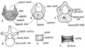 Keith1921 fig056.jpg 1,200 × 681; 150 KB
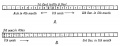 Keith1921 fig057.jpg 1,200 × 459; 72 KB
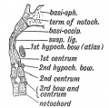 Keith1921 fig058.jpg 623 × 617; 79 KB
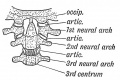 Keith1921 fig059.jpg 759 × 506; 85 KB
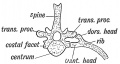 Keith1921 fig060.jpg 809 × 433; 61 KB
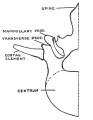 Keith1921 fig061.jpg 520 × 671; 36 KB
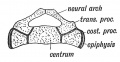 Keith1921 fig062.jpg 602 × 313; 43 KB
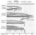 Keith1921 fig063.jpg 948 × 910; 211 KB
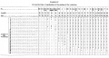 Mall1906 table04.jpg 2,113 × 1,125; 463 KB
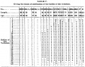 Mall1906 table05.jpg 1,325 × 1,048; 314 KB
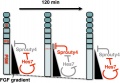 Model for Sprouty4 and FGF in mesoderm segmentation.jpg 481 × 332; 28 KB
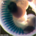 Mouse E11.5 Myog expression.jpg 397 × 397; 25 KB
 Mouse E12.5 Myog Expression.jpg 397 × 397; 17 KB
 Mouse E12.5 Sox9 Expression.jpg 397 × 397; 26 KB
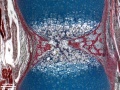 Ossification endochondral 01.jpg 817 × 613; 198 KB
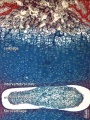 Ossification endochondral 1.jpg 750 × 1,000; 147 KB
 Scoliometry 01.jpg 598 × 449; 38 KB
 Scoliosis drawn.jpg 404 × 1,070; 43 KB
 Scoliosis GH.jpg 600 × 1,053; 87 KB
 Scoliosis xray.jpg 167 × 300; 5 KB
 Scoliosis.jpg 300 × 283; 6 KB
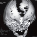 Skull vault defect and midface hypoplasia.jpg 800 × 798; 81 KB
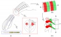 Somite patterning.jpg 600 × 366; 58 KB
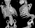 Spina bifida.jpg 800 × 633; 77 KB
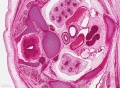 Stage 22 image 200.jpg 1,200 × 877; 563 KB
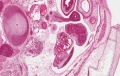 Stage 22 image 202.jpg 1,455 × 920; 617 KB
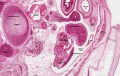 Stage 22 image 302.jpg 1,455 × 920; 625 KB
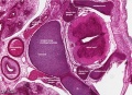 Stage22 vertebra and spinal cord 1.jpg 1,000 × 725; 344 KB
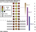 Vertebra ossification placental sequence.jpg 500 × 440; 48 KB
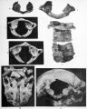 Wheeler1920 fig05-11.jpg 802 × 1,000; 168 KB


