File:Deer mice oocytes at various stages of development in vitro.jpg
Deer_mice_oocytes_at_various_stages_of_development_in_vitro.jpg (686 × 338 pixels, file size: 56 KB, MIME type: image/jpeg)
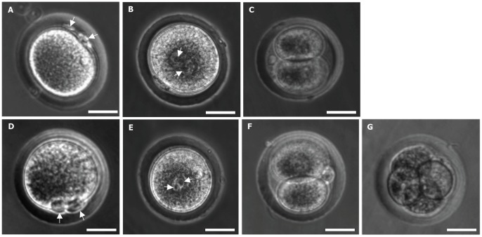
Morphology of in vitro fertilized (IVF) deer mice oocytes at various stages of development in vitro
Typical phase contrast micrographs of developing embryos derived from IVF of MII oocytes retrieved from superovulation (A-C) and IVM (D-G).
Fertilized oocytes with first and secondary polar bodies are clearly visible as indicated by arrows in panels (A) and (D), which was followed by the appearance of two pronuclei (one from oocytes before fertilization and one from sperm) as indicated by arrows in panels (B) and (E) and further development to 2-cell (C and F), and 4-cell (G) stages in vitro.
Scale bar: 30 µm.
Reference
<pubmed>23457518</pubmed>| [http://journals.plos.org/plosone/article?id=10.1371/journal.pone.0056158
Copyright
© 2013 Choi, He. This is an open-access article distributed under the terms of the Creative Commons Attribution License, which permits unrestricted use, distribution, and reproduction in any medium, provided the original author and source are credited.
Figure 3. doi:10.1371/journal.pone.0056158.g003
File history
Yi efo/eka'e gwa ebo wo le nyangagi wuncin ye kamina wunga tinya nan
| Gwalagizhi | Nyangagi | Dimensions | User | Comment | |
|---|---|---|---|---|---|
| current | 16:16, 19 August 2015 | | 686 × 338 (56 KB) | Z3463514 (talk | contribs) | Pone.0056158.g003 PMID 23457518 |
You cannot overwrite this file.
File usage
The following page uses this file: