File:Frazer1926 plate01.jpg
Original file (1,914 × 2,681 pixels, file size: 469 KB, MIME type: image/jpeg)
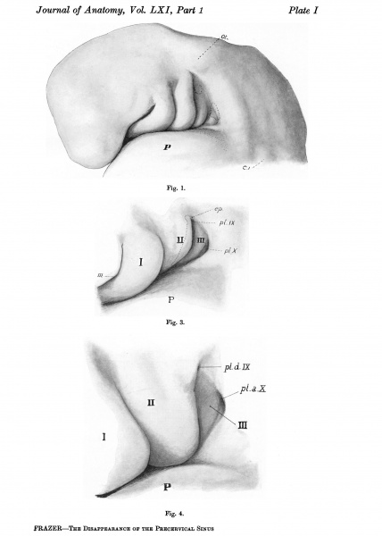
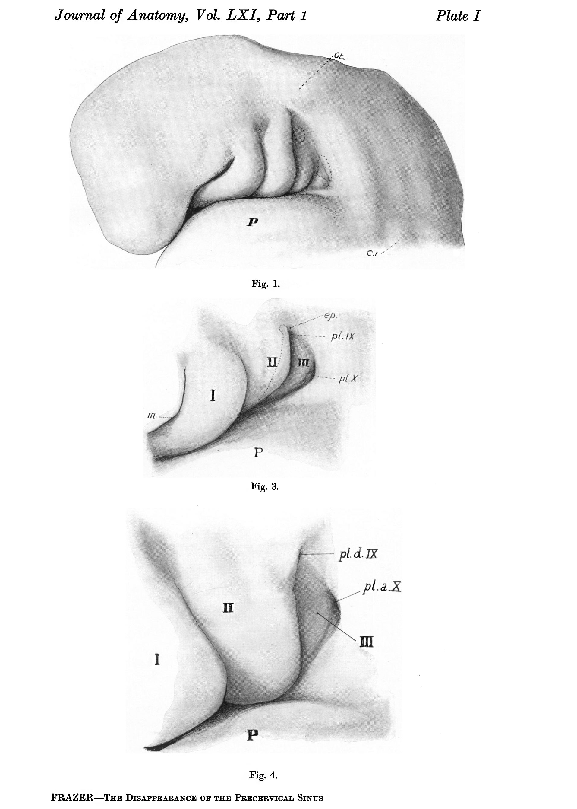
Plate 1.
Fig. 1. Head of embryo of 4-9 mm., seen from the left. P. pericardium; C. 1, first cervical myotome; Ot.,otocyst.
Fig. 3. The precervical sinus in an embryo of 8 mm. seen from the left side. For description see text.
Fig.4. Region of precervical sinus in 10mm. embryo. Left side. P,pericardium;I,II,III, pharyngeal arches. The widely open placodal recess of vagus, pl.a.X, is below and behind the narrow duct, pl.d.IX, leading to the glossopharyngeal ganglion. The epipericardial ridge is very prominent below and behind the third arch.
Cite this page: Hill, M.A. (2026, October 5) Embryology Frazer1926 plate01.jpg. Retrieved from https://embryology.med.unsw.edu.au/embryology/index.php/File:Frazer1926_plate01.jpg
- © Dr Mark Hill 2026, UNSW Embryology ISBN: 978 0 7334 2609 4 - UNSW CRICOS Provider Code No. 00098G
File history
Yi efo/eka'e gwa ebo wo le nyangagi wuncin ye kamina wunga tinya nan
| Gwalagizhi | Nyangagi | Dimensions | User | Comment | |
|---|---|---|---|---|---|
| current | 11:28, 28 July 2015 | | 1,914 × 2,681 (469 KB) | Z8600021 (talk | contribs) |
You cannot overwrite this file.
File usage
The following page uses this file: