File:Anson1934 plate02.jpg

Original file (1,464 × 2,311 pixels, file size: 259 KB, MIME type: image/jpeg)
Plate 2 Auditory Vesicle Models from Harvard Embryological Collection
| Historic Disclaimer - information about historic embryology pages |
|---|
 |
- Links: fig 1-7 | fig 1 | fig 2 | fig 3 | fig 4 | fig 5 | fig 6 | fig 7 | fig 8-21 | fig 22-33 | 1934 Anson | Historic Papers | Inner Ear Development
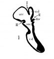
fig 1 Human 22.8 mm
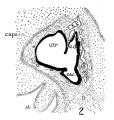
fig 2 Human 29 mm
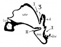
fig 3 Human 34 mm
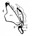
fig 4 Human 40 mm
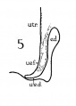
fig 5 Infant 4 months
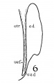
fig 6 Child 3 years
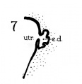
fig 7 embryo 36 mm
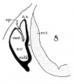
8 Cat 7 mm
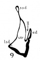
9 Cat 10.6 mm
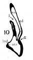
10 Cat 14 mm
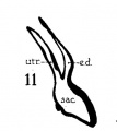
11 Cat 15 mm
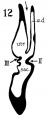
12 Cat 24.1 mm
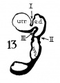
13 Cat 32.6 mm
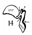
14 Cat 39 mm
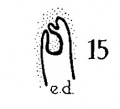
15 Cat 39 mm
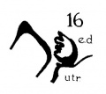
16 Cat 31 mm
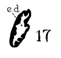
17 Guinea-pig 18.5 mm
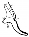
18 Dog 12.5 mm
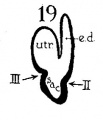
19 Rabbit 21 mm
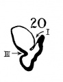
20 Rabbit 25 mm
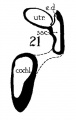
21 Rabbit 29 mm


Reference
Anson BJ. The early development of the membranous labyrinth in mammalian embryos, with special reference to the endolymphatic duct and the utriculo—endolymphatic duct. (1934) Anat. Rec. 59: 15-25.
Cite this page: Hill, M.A. (2026, October 3) Embryology Anson1934 plate02.jpg. Retrieved from https://embryology.med.unsw.edu.au/embryology/index.php/File:Anson1934_plate02.jpg
- © Dr Mark Hill 2026, UNSW Embryology ISBN: 978 0 7334 2609 4 - UNSW CRICOS Provider Code No. 00098G
File history
Yi efo/eka'e gwa ebo wo le nyangagi wuncin ye kamina wunga tinya nan
| Gwalagizhi | Nyangagi | Dimensions | User | Comment | |
|---|---|---|---|---|---|
| current | 15:45, 23 July 2015 | | 1,464 × 2,311 (259 KB) | Z8600021 (talk | contribs) |
You cannot overwrite this file.
File usage
The following page uses this file:


