File:Hill1931 textfig02.jpg
From Embryology
Size of this preview: 536 × 599 pixels. Other resolution: 1,413 × 1,580 pixels.
Original file (1,413 × 1,580 pixels, file size: 171 KB, MIME type: image/jpeg)
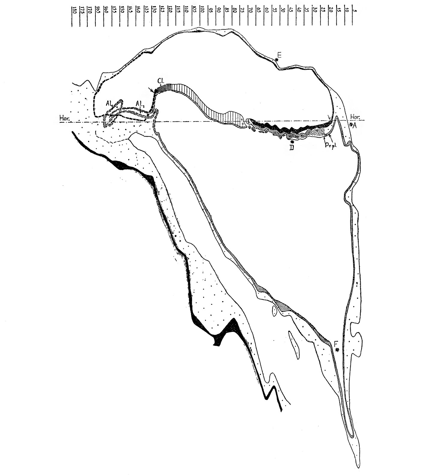
Text-Fig. 2. Graphic reconstruction of the median section through the Dobbin embryo
original figure x72
Ectoderm black; endoderm lined horizontally; primitive streak lined vertically; + = Hensen's knot; chords-process lined obliquely; prochordal plate (Pr.Pl.) lined horizontally and dotted; mesoderm dotted; cloacal membrane (Cl.) cross-hatched.
- Links: Text-fig. 1 | Text-fig. 2 | Text-fig. 3 | Text-fig. 4 | Text-fig. 5 | Text-fig. 6 | Text-fig. 7 | Text-fig. 8 | Text-fig. 9 | Text-fig. 10 | Text-fig. 11 | Text-fig. 12 | Text-fig. 13 | Text-fig. 14 | Text-fig. 15 | Text-fig. 16 | Text-fig. 17 | Primitive streak table | Plate 29 | Plate 30 | Plate 31 | Plate 32 | Plate 33
Reference
Hill JP., Florian J, A Young Human Embryo (Embryo Dobbin) with Head-process nd Prechordal Plate. (1931) Philosophical Transactions of the Royal Society of London. Series B, Containing Papers of a Biological Character.
File history
Click on a date/time to view the file as it appeared at that time.
| Date/Time | Thumbnail | Dimensions | User | Comment | |
|---|---|---|---|---|---|
| current | 19:15, 12 April 2015 | | 1,413 × 1,580 (171 KB) | Z8600021 (talk | contribs) | |
| 19:14, 12 April 2015 | 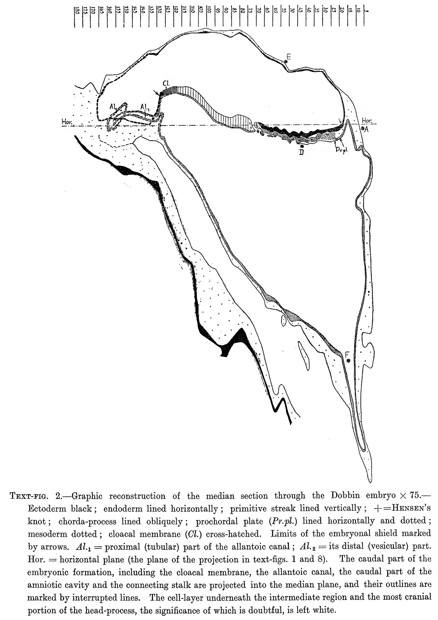 | 1,414 × 2,000 (335 KB) | Z8600021 (talk | contribs) |
You cannot overwrite this file.
File usage
There are no pages that use this file.