File:Florian1935 textfig01.jpg

Original file (1,120 × 1,202 pixels, file size: 136 KB, MIME type: image/jpeg)
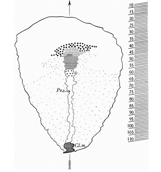
Text fig.1. Graphic reconstruction of the dorsal view of the embryonal shield
x100.
The outline of the embryonal shield marked by a thick line. Outline of the primitive streak (Fr.s.) and Heunsen's knot marked by a dotted line; Heusen's knot marked by crosses. Cloacal membrane (C'l.m.) cross-hatched. Head-process marked by horizontal lines closely arranged in its compact, thicker caudal part (chorda process), more widely spaced with dots between, in its wider and more loosely constituted cranial part. prochordal plate marked by large dots. Secondary mesoderm below The embryonlal shield marked by by fine dots. On the right of the figure, levels of sections 8-110 are indicated. Median plane marked by an arrow.
File history
Yi efo/eka'e gwa ebo wo le nyangagi wuncin ye kamina wunga tinya nan
| Gwalagizhi | Nyangagi | Dimensions | User | Comment | |
|---|---|---|---|---|---|
| current | 13:50, 12 April 2015 | | 1,120 × 1,202 (136 KB) | Z8600021 (talk | contribs) | |
| 13:49, 12 April 2015 | 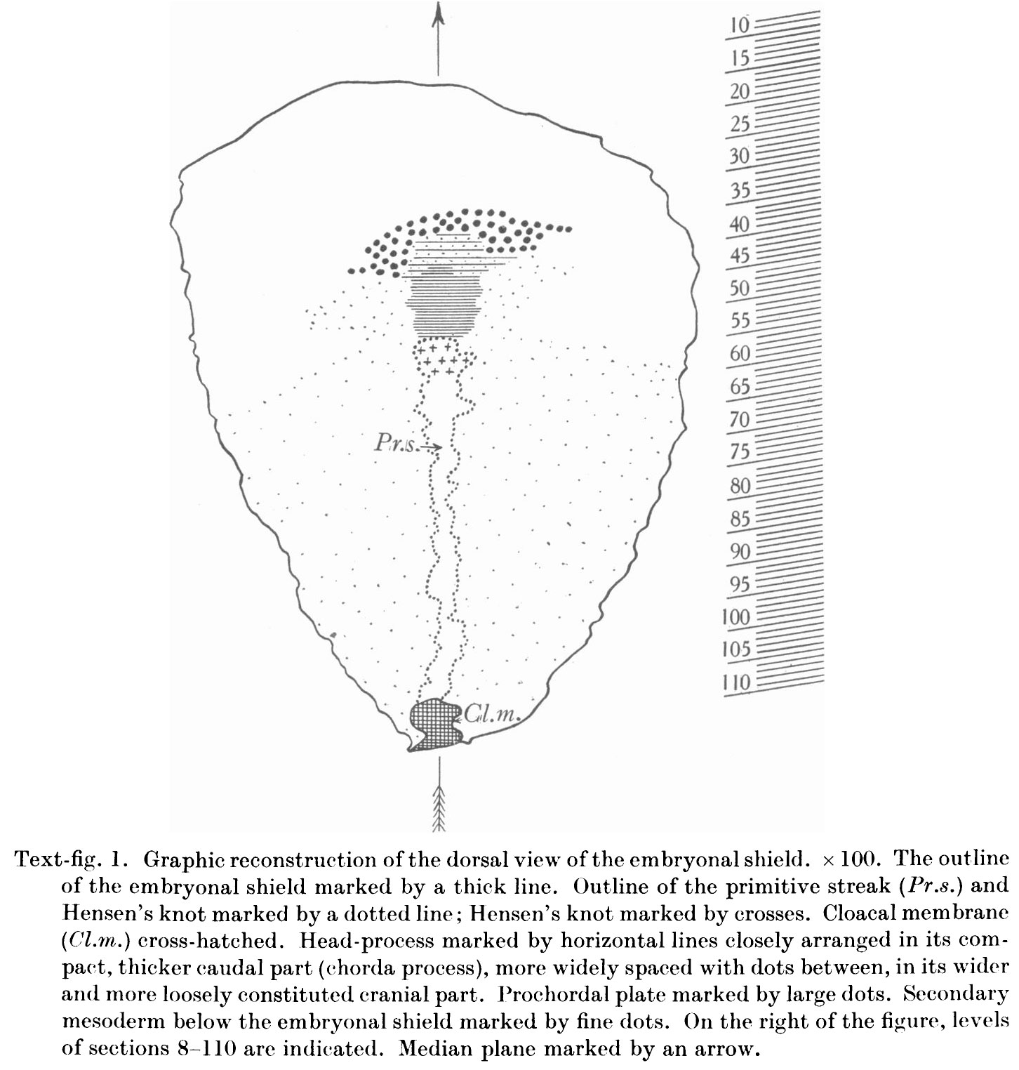 | 1,452 × 1,524 (257 KB) | Z8600021 (talk | contribs) |
You cannot overwrite this file.
File usage
The following 4 pages use this file: