File:Minot1897 005.jpg
From Embryology

Size of this preview: 800 × 493 pixels. Other resolution: 953 × 587 pixels.
Original file (953 × 587 pixels, file size: 132 KB, MIME type: image/jpeg)
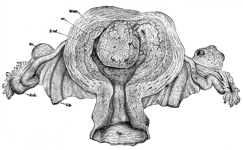
Fig. 5. Uterus about forty days advanced in pregnancy
after Conte.
The uterus has been opened by cutting through the anterior wall and reflecting the sides.
- Musc. - muscularis
- De. - decidua vera
- D ref. - decidua reflexa
- Ov. - ovary
- Ovd. - Fallopian tube
- Lig. - round ligament
- Vg. - vagina
| Historic Disclaimer - information about historic embryology pages |
|---|
 |
File history
Yi efo/eka'e gwa ebo wo le nyangagi wuncin ye kamina wunga tinya nan
| Gwalagizhi | Nyangagi | Dimensions | User | Comment | |
|---|---|---|---|---|---|
| current | 21:04, 1 April 2014 | | 953 × 587 (132 KB) | Z8600021 (talk | contribs) | {{Historic Disclaimer}} Category: Charles Minot |
You cannot overwrite this file.
File usage
The following 2 pages use this file: