File:Fetal week 14 head bone lateral 01.jpg
From Embryology
Size of this preview: 776 × 600 pixels. Other resolution: 1,000 × 773 pixels.
Original file (1,000 × 773 pixels, file size: 107 KB, MIME type: image/jpeg)
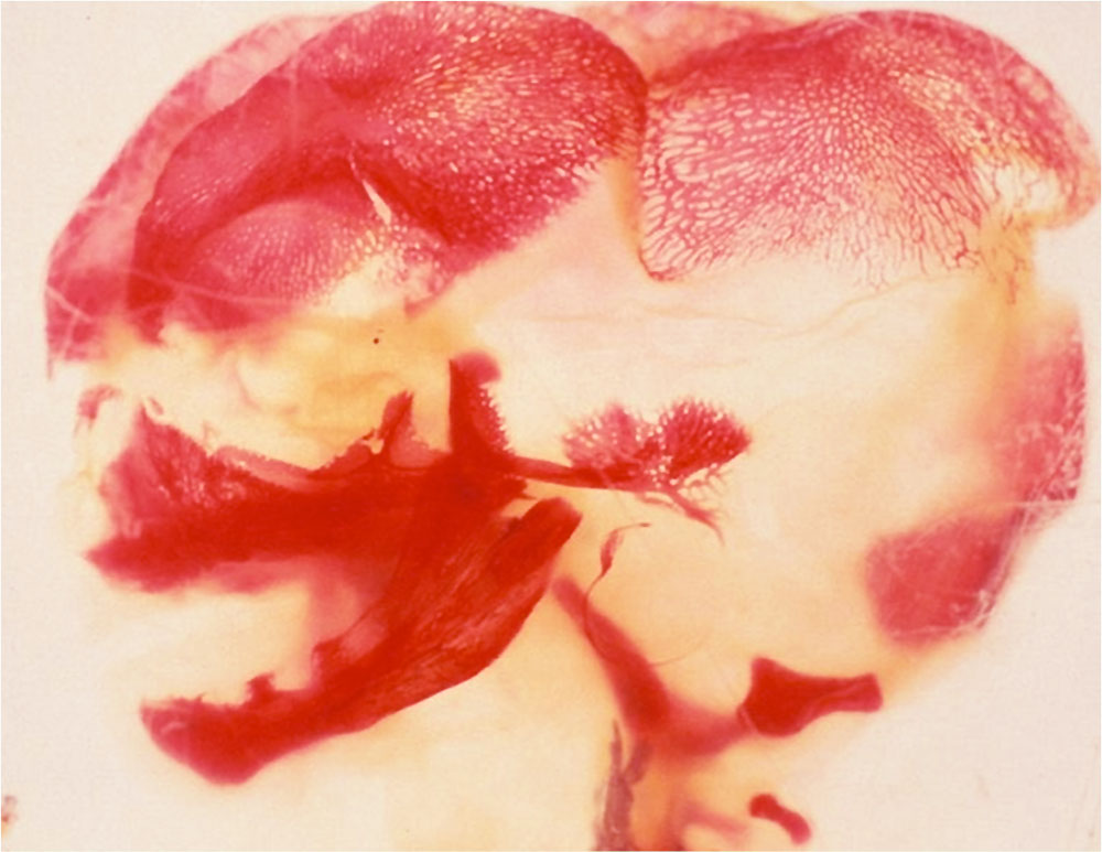
Fetal Head (14 weeks) Lateral View
Fetal Skull has been stained to show regions of bone development (ossification).
Image Source: Prof Virginia Diewert
Cite this page: Hill, M.A. (2024, June 27) Embryology Fetal week 14 head bone lateral 01.jpg. Retrieved from https://embryology.med.unsw.edu.au/embryology/index.php/File:Fetal_week_14_head_bone_lateral_01.jpg
- © Dr Mark Hill 2024, UNSW Embryology ISBN: 978 0 7334 2609 4 - UNSW CRICOS Provider Code No. 00098G
File history
Yi efo/eka'e gwa ebo wo le nyangagi wuncin ye kamina wunga tinya nan
| Gwalagizhi | Nyangagi | Dimensions | User | Comment | |
|---|---|---|---|---|---|
| current | 14:33, 15 May 2013 | | 1,000 × 773 (107 KB) | Z8600021 (talk | contribs) | ==Fetal Head (14 weeks) Lateral View== ---- Image Source: Prof Virginia Diewert {{Template:Footer}} Category:Human Fetus Category:Musculoskeletal Category:Bone Category:Head Category:Week 14 Category:Skull |
You cannot overwrite this file.
File usage
The following page uses this file: