File:Gray0617.jpg
Gray0617.jpg (555 × 600 pixels, file size: 139 KB, MIME type: image/jpeg)
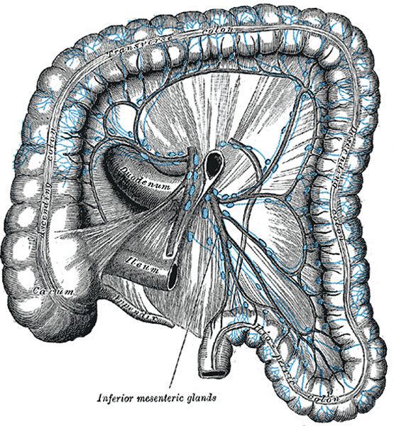
Lymphatics of Colon
(Jamieson and Dobson.)
Mesocolic Glands (lymphoglandulæ mesocolicæ) are numerous, and lie between the layers of the transverse mesocolon, in close relation to the transverse colon; they are best developed in the neighborhood of the right and left colic flexures. One or two small glands are occasionally seen along the trunk of the right colic artery and others are found in relation to the trunk and branches of the middle colic artery.
Superior mesenteric glands receive afferents from the jejunum, ileum, cecum, vermiform process, and the ascending and transverse parts of the colon; their efferents pass to the preaortic glands.
Inferior mesenteric glands (Fig. 617) consist of:
- small glands on the branches of the left colic and sigmoid arteries
- a group in the sigmoid mesocolon, around the superior hemorrhoidal artery
- a pararectal group in contact with the muscular coat of the rectum.
They drain the descending iliac and sigmoid parts of the colon and the upper part of the rectum; their efferents pass to the preaortic glands.
(Text from Gray's Anatomy 1918)
- Gray's Images: Development | Lymphatic | Neural | Vision | Hearing | Somatosensory | Integumentary | Respiratory | Gastrointestinal | Urogenital | Endocrine | Surface Anatomy | iBook | Historic Disclaimer
| Historic Disclaimer - information about historic embryology pages |
|---|
 |
| iBook - Gray's Embryology | |
|---|---|

|
|
Reference
Gray H. Anatomy of the human body. (1918) Philadelphia: Lea & Febiger.
Cite this page: Hill, M.A. (2024, June 11) Embryology Gray0617.jpg. Retrieved from https://embryology.med.unsw.edu.au/embryology/index.php/File:Gray0617.jpg
- © Dr Mark Hill 2024, UNSW Embryology ISBN: 978 0 7334 2609 4 - UNSW CRICOS Provider Code No. 00098G
File history
Click on a date/time to view the file as it appeared at that time.
| Date/Time | Thumbnail | Dimensions | User | Comment | |
|---|---|---|---|---|---|
| current | 23:55, 14 February 2013 | | 555 × 600 (139 KB) | Z8600021 (talk | contribs) | (Text from Gray's Anatomy 1918) {{Gray Anatomy}} Category:Immune |
You cannot overwrite this file.
File usage
The following 3 pages use this file: