File:Formation of the eyelid 2.jpg
From Embryology
Size of this preview: 800 × 450 pixels. Other resolution: 1,152 × 648 pixels.
Original file (1,152 × 648 pixels, file size: 78 KB, MIME type: image/jpeg)
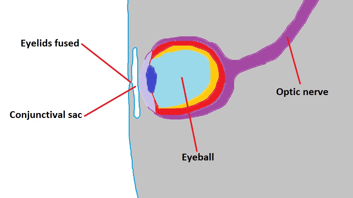
A schematic showing the eye at an advanced stage of embryonic development. Note that all major components are now in place, however the eyelids remain fused until several weeks later.
Copyright: This is a student drawn image and is free for non-profit reuse.
--Mark Hill 00:37, 3 October 2012 (EST) There is no timing information provided here. This simple diagram requires more information to be useful in your project.
- Note - This image was originally uploaded as part of an undergraduate science student project and may contain inaccuracies in either description or acknowledgements. Students have been advised in writing concerning the reuse of content and may accidentally have misunderstood the original terms of use. If image reuse on this non-commercial educational site infringes your existing copyright, please contact the site editor for immediate removal.
File history
Yi efo/eka'e gwa ebo wo le nyangagi wuncin ye kamina wunga tinya nan
| Gwalagizhi | Nyangagi | Dimensions | User | Comment | |
|---|---|---|---|---|---|
| current | 08:32, 2 October 2012 | | 1,152 × 648 (78 KB) | Z3373894 (talk | contribs) | A schematic showing the eye at an advanced stage of embryonic development. Note that all major components are now in place, however the eyelids remain fused until several weeks later. Copyright: This is a student drawn image and is free for non-profit re |
You cannot overwrite this file.
File usage
The following 2 pages use this file: