File:Week 10 Midgut Herniation.png
Original file (865 × 603 pixels, file size: 505 KB, MIME type: image/png)
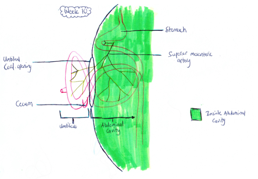
Week 10 Herniated Midgut
This is a hand drawn of the week 10 fetal midgut. It is based off the images from Martin V, Shaw-Smith C, [1] The Midgut which was initially herniated in week 5 of gestation is still lying outside the abdominal cavity (shaded green) in the umbilicus. This is shown by the red loop of midgut which are the intestinal loops protruding through the abdominal cavity. This article focuses on midgut malrotation, however in doing so describes the embryonic rotations that occur and the herniation of the midgut to get it to the position illustrated in this diagram.
References
- ↑ name="PMID2908440"><pubmed>2908440</pubmed>
--Mark Hill (talk) 11:01, 7 November 2014 (EST) Assessment - Student drawn image. Figure relates to project topic contains reference, copyright and student template. Caption information is useful. The drawing does show herniation but it is not clearly illustrated in your drawing. Minor, there is a problem with your reference formatting.
- Note - This image was originally uploaded as part of an undergraduate science student project and may contain inaccuracies in either description or acknowledgements. Students have been advised in writing concerning the reuse of content and may accidentally have misunderstood the original terms of use. If image reuse on this non-commercial educational site infringes your existing copyright, please contact the site editor for immediate removal.
File history
Yi efo/eka'e gwa ebo wo le nyangagi wuncin ye kamina wunga tinya nan
| Gwalagizhi | Nyangagi | Dimensions | User | Comment | |
|---|---|---|---|---|---|
| 22:28, 21 October 2014 |  | 865 × 605 (503 KB) | Z3415141 (talk | contribs) | ||
| 22:27, 21 October 2014 |  | 865 × 605 (503 KB) | Z3415141 (talk | contribs) | Reverted to version as of 12:21, 21 October 2014 | |
| 22:26, 21 October 2014 |  | 865 × 605 (503 KB) | Z3415141 (talk | contribs) | Reverted to version as of 12:22, 21 October 2014: After receiving feedback from my peers about the colour of the labels I have now changed the labels to being black so that it they are more clear and easier to read. | |
| 22:25, 21 October 2014 | 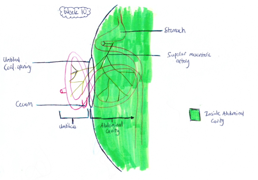 | 865 × 605 (503 KB) | Z3415141 (talk | contribs) | Reverted to version as of 12:21, 21 October 2014 | |
| 22:22, 21 October 2014 |  | 865 × 605 (503 KB) | Z3415141 (talk | contribs) | After receiving feedback from my peers about the colour of the labels I have now changed the labels to being black so that it they are more clear and easier to read. | |
| 22:21, 21 October 2014 |  | 865 × 605 (503 KB) | Z3415141 (talk | contribs) | After receiving feedback from my peers about the colour of the labels I have now changed the labels to being black so that it they are more clear and easier to read. | |
| 19:16, 6 October 2014 | 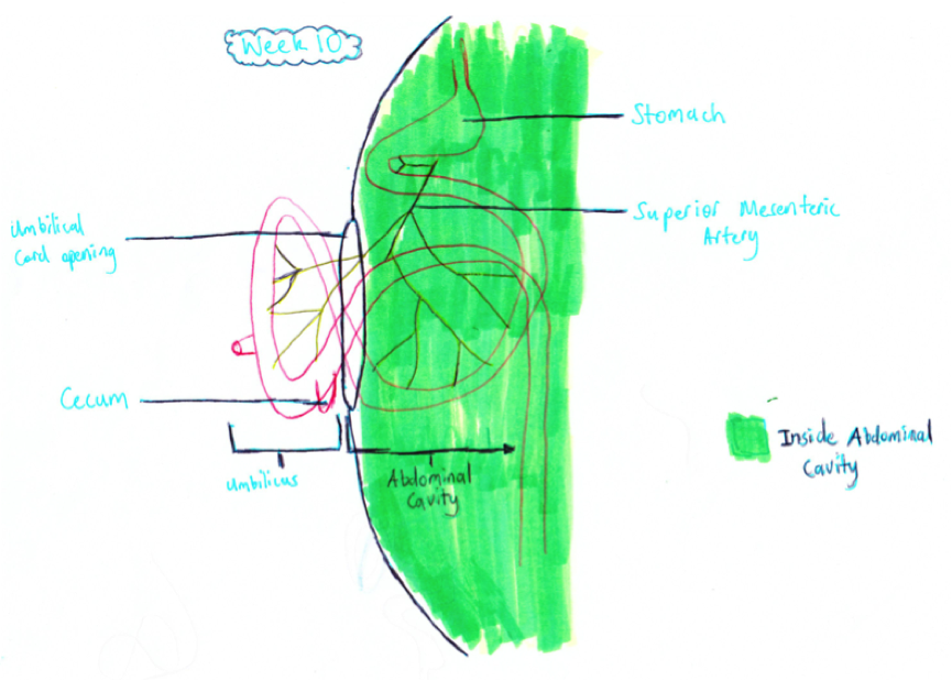 | 865 × 619 (538 KB) | Z3415141 (talk | contribs) | ||
| 18:49, 6 October 2014 | 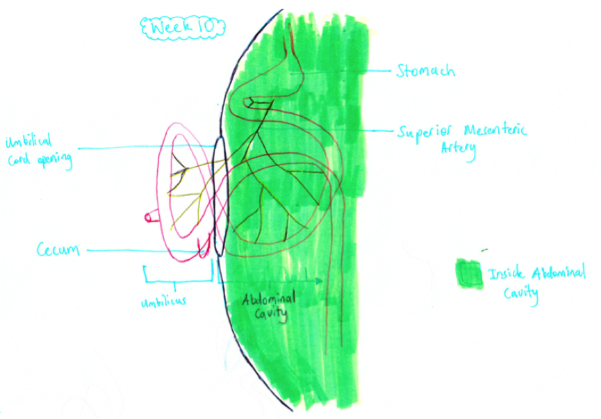 | 865 × 603 (512 KB) | Z3415141 (talk | contribs) | Reverted to version as of 08:40, 6 October 2014 | |
| 18:48, 6 October 2014 |  | 865 × 603 (512 KB) | Z3415141 (talk | contribs) | Week 10 Fetal midgut: Lying in the umbilicus outside the green part of this image is the herniated midgut<ref name="PMID2908440"><pubmed>2908440</pubmed></ref>. | |
| 18:40, 6 October 2014 |  | 865 × 603 (512 KB) | Z3415141 (talk | contribs) | Week 10 Fetal midgut: Lying in the umbilicus outside the green part of this image is the herniated midgut. |
You cannot overwrite this file.
File usage
The following 2 pages use this file: