File list
From Embryology
This special page shows all uploaded files.
| Date | Name | Thumbnail | Size | User | Description | Versions |
|---|---|---|---|---|---|---|
| 11:22, 14 March 2010 | Aortic Stenosis.jpg (file) |  |
16 KB | Z3212774 | category:Heart ILP | 1 |
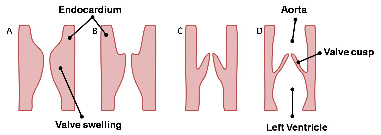
| 11:17, 14 March 2010 | Semilunar Cusps.jpg (file) |  |
52 KB | Z3212774 | category:Heart ILP Longitudinal sections of the aorta showing development of the semilunar cusps forming the aortic valve. | 1 |
| 11:16, 14 March 2010 | Semilunar Valves.jpg (file) |  |
93 KB | Z3212774 | category:Heart ILP Development of the semilunar valves of the aorta and pulmonary trunk as shown through a transverse section of the bulbus cordis. The walls and valves of the aorta and pulmonary trunk form followed by rotation of the vessels which e | 1 |
| 11:15, 14 March 2010 | AV Valves.jpg (file) |  |
129 KB | Z3212774 | category:Heart ILP Sequence of events in the development of the atrioventricular valves. The structures of the valves i.e. the papillary muscles, chordae tendineae and cusps are sculpted from the muscular ventricular walls. | 1 |
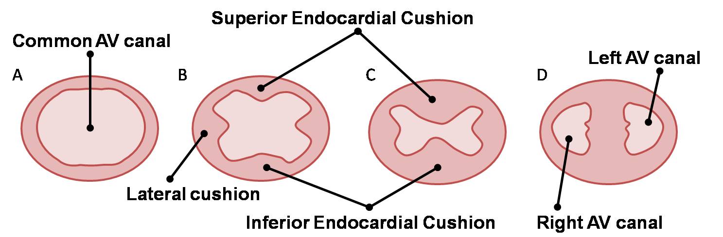
| 11:14, 14 March 2010 | AV Canal Division (Superior View).jpg (file) | .jpg) |
69 KB | Z3212774 | category:Heart ILP Development of the atrioventricular septum over weeks four and five. The right and left atrioventricular canals are remodelled to later become the atrioventricular (tricuspid and mitral) valves. | 1 |
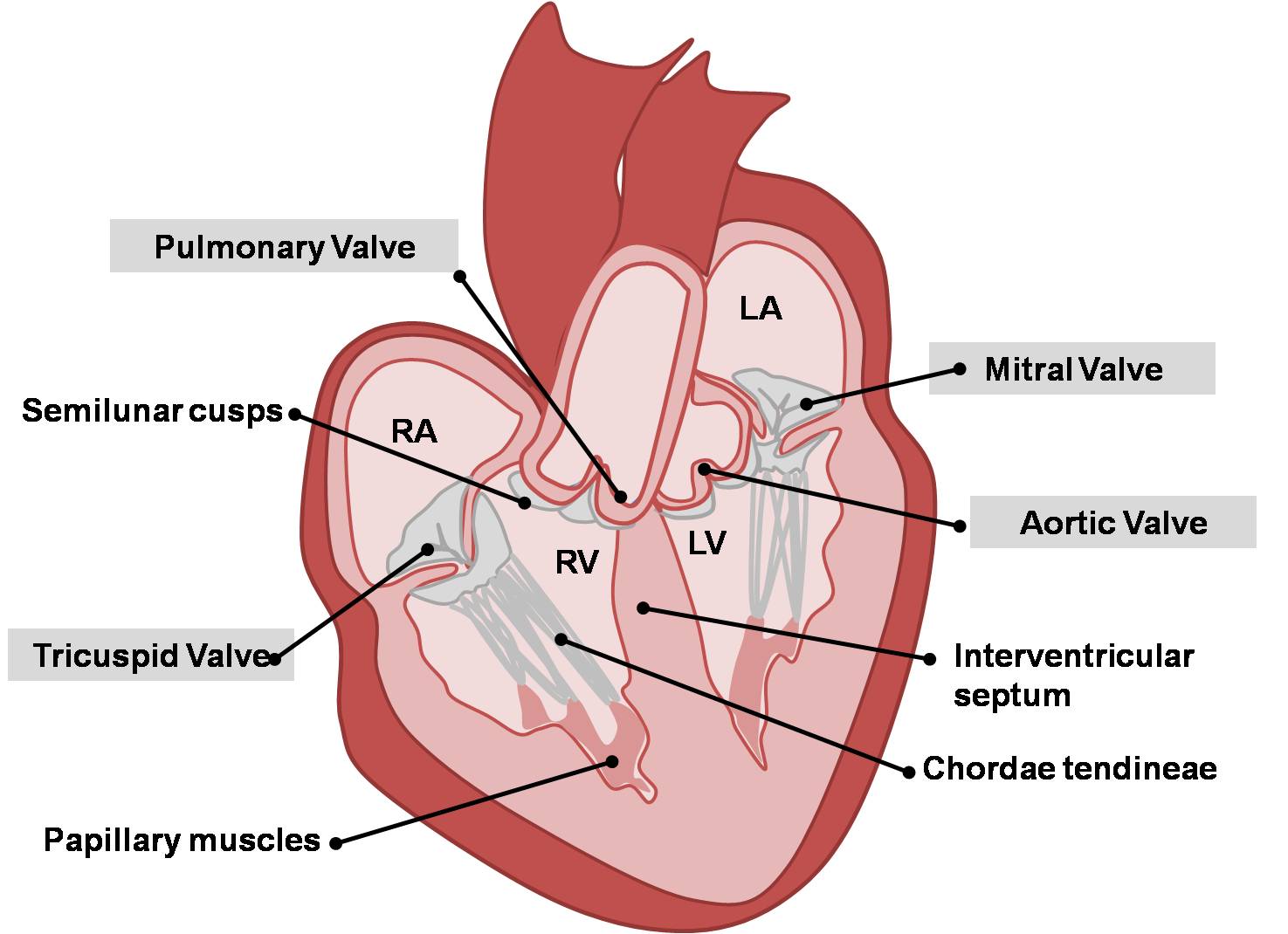
| 11:13, 14 March 2010 | Adult Heart Valves.jpg (file) |  |
113 KB | Z3212774 | category:Heart ILP Adult heart showing aortic, pulmonary, mitral and tricuspid valves. | 1 |
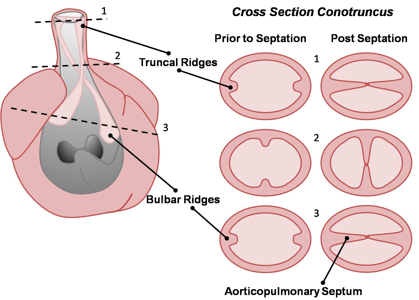
| 11:02, 14 March 2010 | Outflow Tract Division (Cross-Section).jpg (file) | .jpg) |
119 KB | Z3212774 | category:Heart ILP Cross sections of the outflow tract before and after fusion of the conotruncal ridges. | 1 |
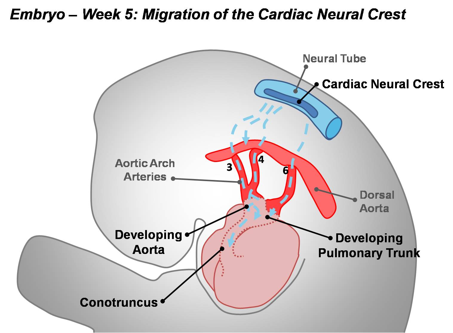
| 11:00, 14 March 2010 | Cardiac Neural Crest Migration.jpg (file) |  |
122 KB | Z3212774 | category:Heart ILP In order to complete division of the outflow tract, mesenchyme derived from the cardiac neural crest migrates over the aortic arch arteries to invade the conotruncus. | 1 |
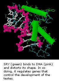
| 10:57, 14 March 2010 | SRY and DNA.jpg (file) |  |
19 KB | S8600021 | Image Source: Genes and Disease NCBI | 1 |
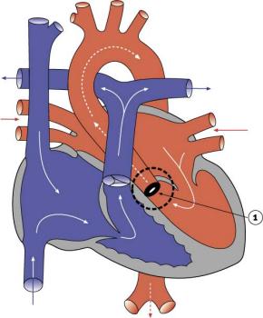
| 10:56, 14 March 2010 | Embryonic Heart Blood Flow.jpg (file) |  |
101 KB | Z3212774 | category:Heart ILP Oxygenated (from the placenta) and non-oxygenated (from the lower body) blood enters the right atrium via the sinus venosus. Part of this blood travels to the right ventricle and then through the pulmonary circulation. The rest of | 1 |
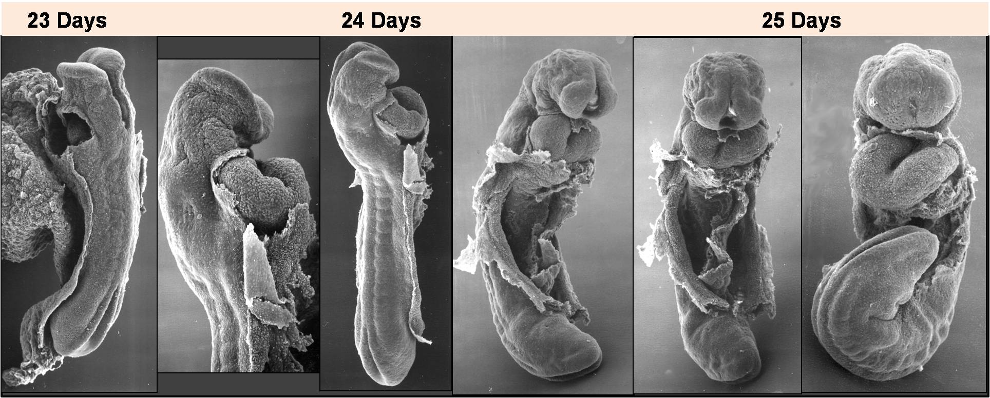
| 10:50, 14 March 2010 | Heart Looping Sequence (SEMs).jpg (file) | .jpg) |
212 KB | Z3212774 | category:Heart ILP {{Template:SEM}} The heart tube loops initially to form a C-shape and as looping progresses the heart begins to resemble an S-shape (or U-shape ventrally). | 1 |
| 10:49, 14 March 2010 | Heart Looping Sequence.jpg (file) |  |
85 KB | Z3212774 | category:Heart ILP Shows the sequence of events in heart looping. The heart begins as a straight tube then bends ventrally. Rotation brings the bulge of the ventral bend (predominantly the bulbus cordis and ventricle) to the right, forming a C-shaped | 1 |
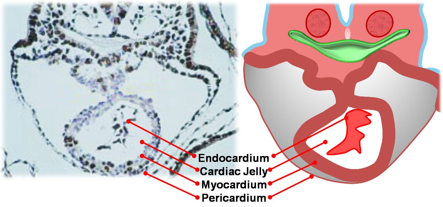
| 10:44, 14 March 2010 | Heart Tube (Cross-Section).jpg (file) | .jpg) |
101 KB | Z3212774 | category:Heart ILP Cross section through the ventricular portion of the heart tube, suspended from the dorsal wall by the dorsal mesocardium. Shows the layers of the heart tube. | 1 |
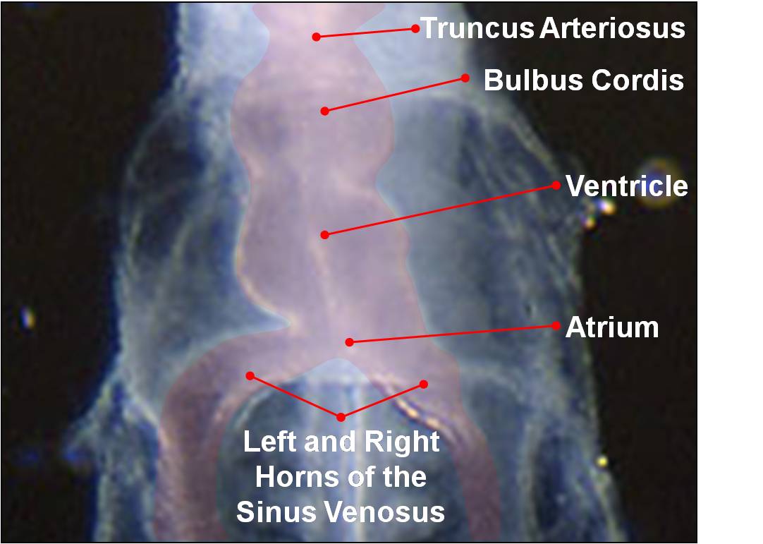
| 10:42, 14 March 2010 | Heart Tube Segments.jpg (file) |  |
63 KB | Z3212774 | category:Heart ILP {{Template:SEM}} As the tubular heart grows it develops dilations and constrictions which form the truncus arteriosus, bulbus cordis, primitive ventricle, primitive atrium and sinus venosus. | 1 |
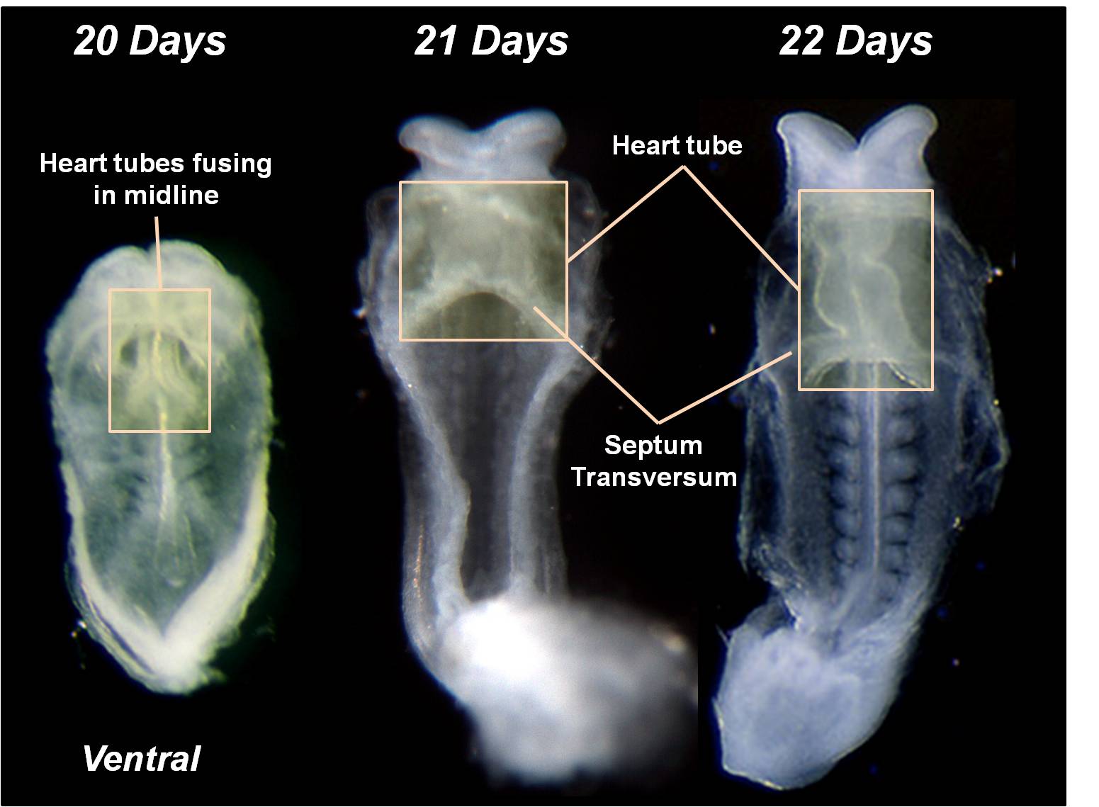
| 10:41, 14 March 2010 | Heart Tube Fusion.jpg (file) |  |
125 KB | Z3212774 | category:Heart ILP {{Template:SEM}} The primordial heart tubes fuse in the midline to form a single ventral heart tube. Fusion begins cranially and extends caudally. | 1 |
| 10:38, 14 March 2010 | Early Heart Tube (Lateral).jpg (file) | .jpg) |
110 KB | Z3212774 | category:Heart ILP Angiogenesis throughout the embryo allows for the development of angioblastic cords in the cardiogenic mesoderm of the embryo. | 1 |
| 10:37, 14 March 2010 | Early Heart Tube (Dorsal).jpg (file) | .jpg) |
111 KB | Z3212774 | category:Heart ILP Angiogenesis throughout the embryo allows for the development of angioblastic cords in the cardiogenic mesoderm of the embryo. | 1 |
| 10:29, 14 March 2010 | Fetal Circulation Pathway.jpg (file) |  |
112 KB | Z3212774 | category:Heart ILP | 1 |
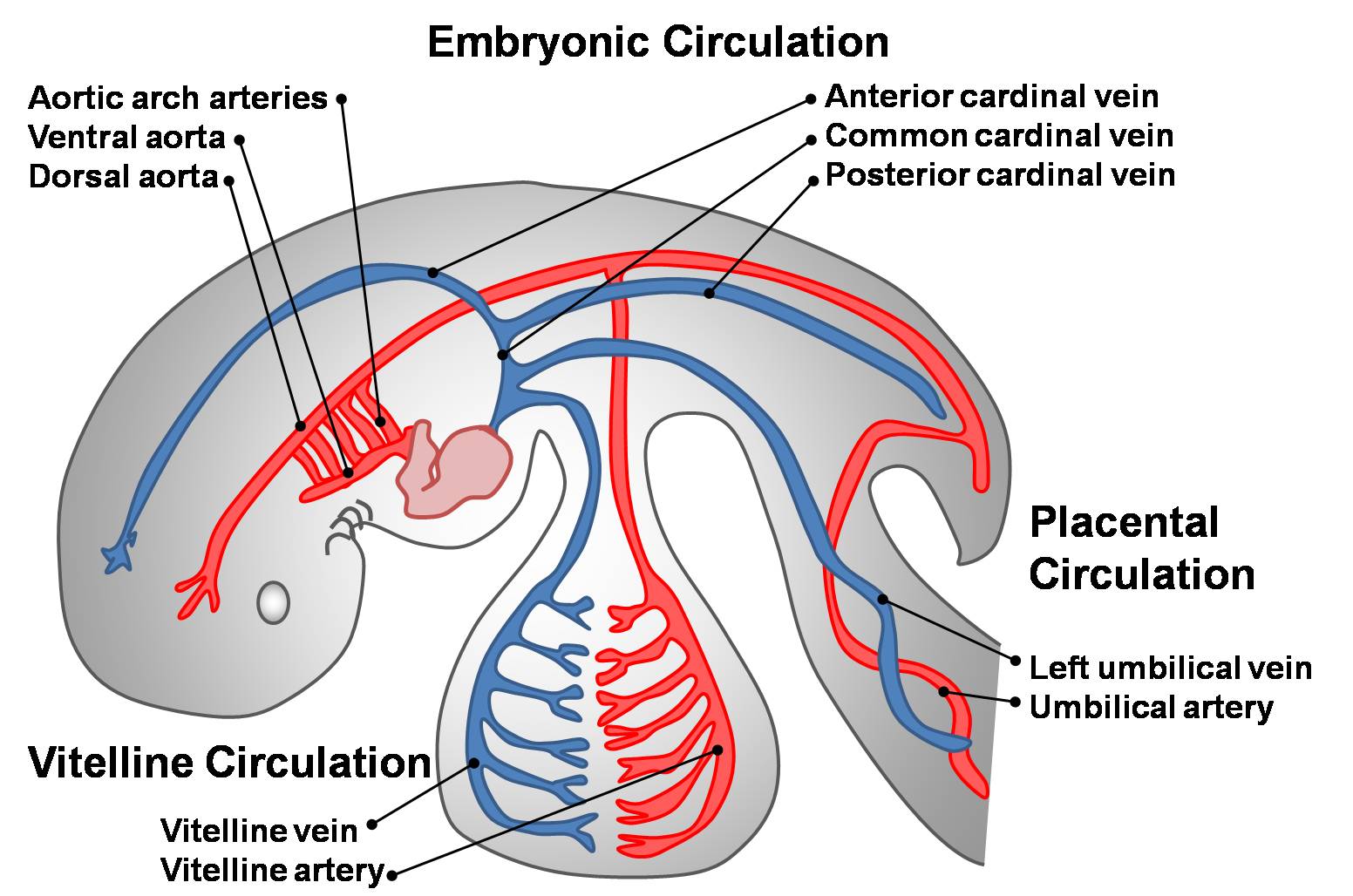
| 10:28, 14 March 2010 | Embryonic Circulations.jpg (file) |  |
171 KB | Z3212774 | category:Heart ILP The three early embryonic circulations. Three paired veins drain into the primordial heart tube: vitelline veins (returning poorly oxygenated blood from the yolk sac), umbilical veins (carrying well-oxygenated blood from the primor | 1 |
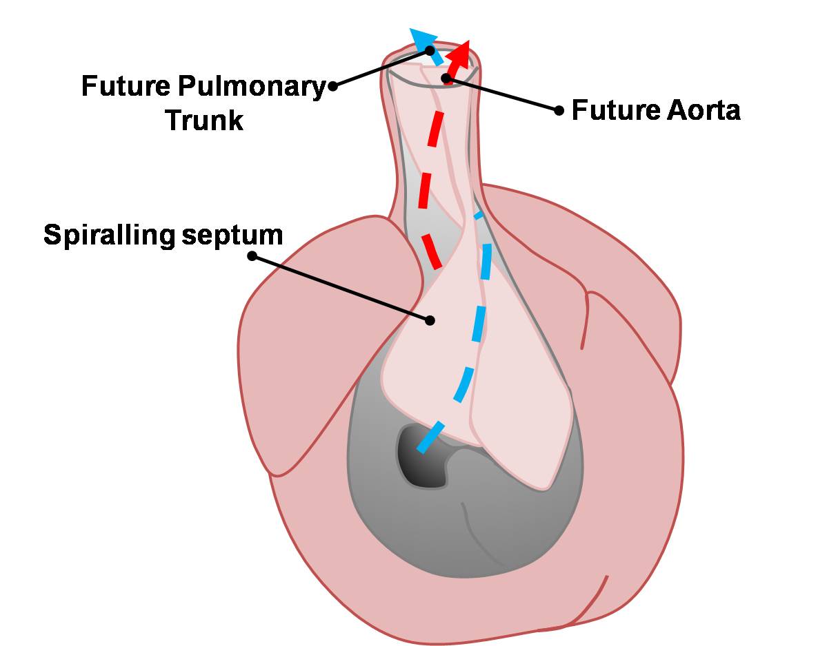
| 10:22, 14 March 2010 | Basic Outflow Tract Division.jpg (file) |  |
59 KB | Z3212774 | category:Heart ILP Blood flow through the conotruncus is divided to form the pulmonary trunk and aorta. | 1 |
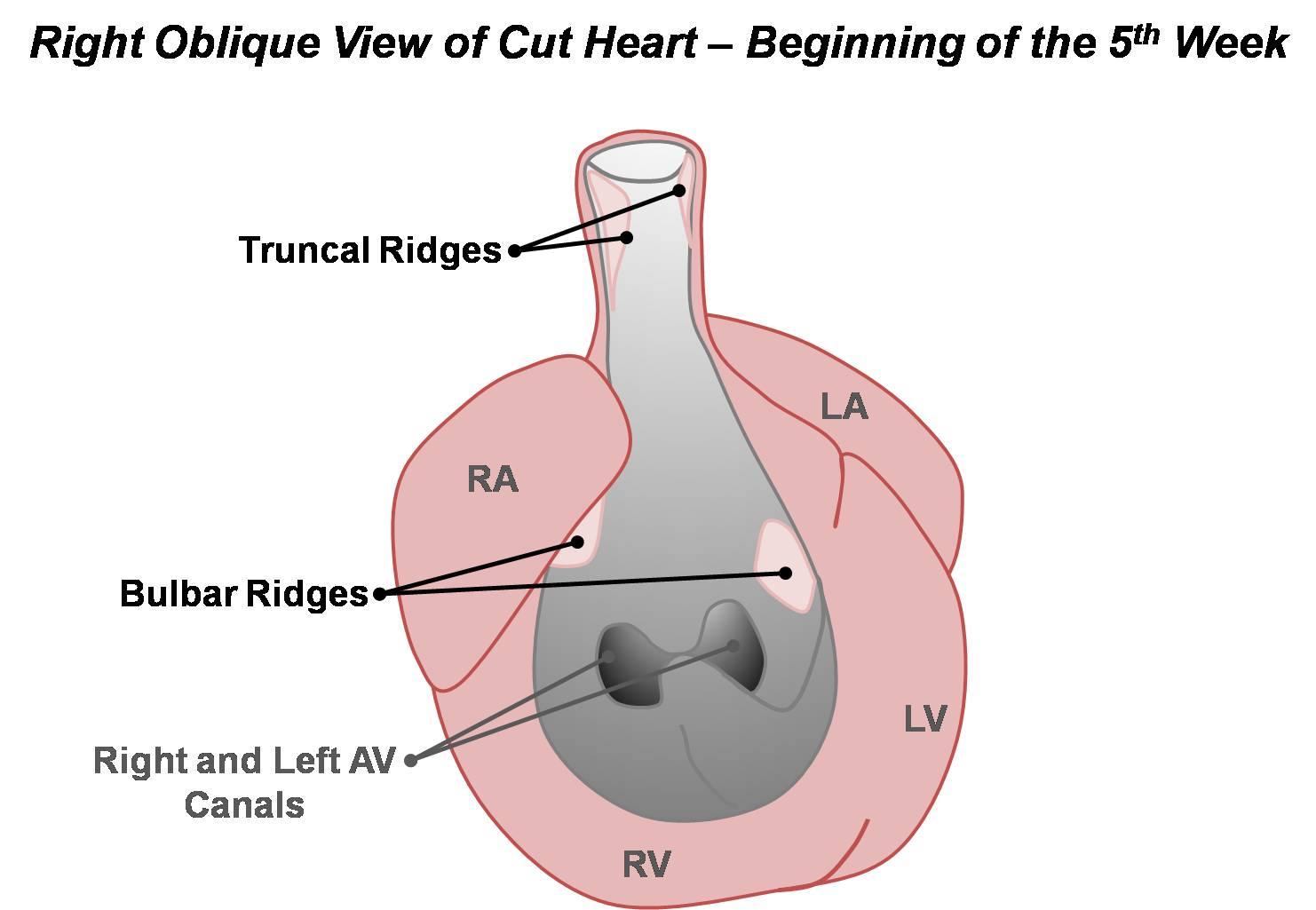
| 10:21, 14 March 2010 | Basic Conotruncal Ridge Development.jpg (file) |  |
89 KB | Z3212774 | category:Heart ILP Truncal and bulbar ridges develop marking the beginning of the division of the outflow tract. | 1 |
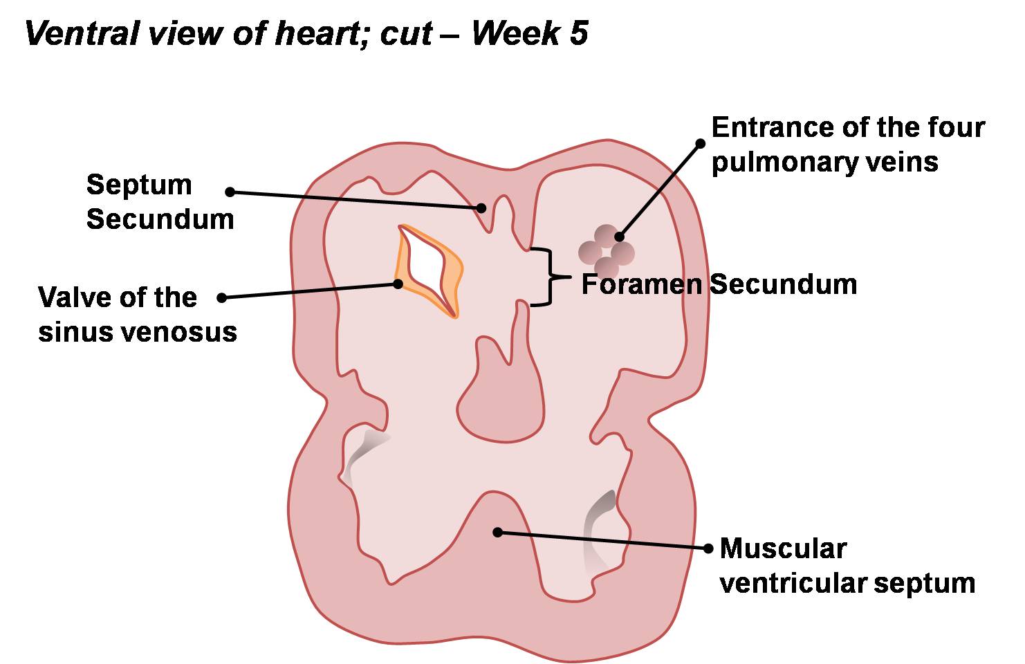
| 10:20, 14 March 2010 | Atrial & Ventricular Septation 2.jpg (file) |  |
113 KB | Z3212774 | category:Heart ILP The embryonic heart begins to resemble the adult heart as septation is complete. A right-to-left shunt exists between the right and left atria via the foramen ovale. Both muscular and membranous portions of the ventricular septum a | 1 |
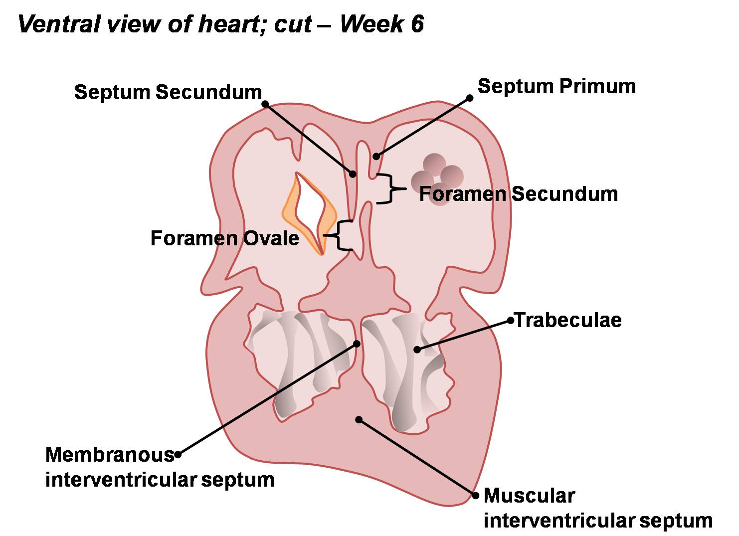
| 10:20, 14 March 2010 | Atrial & Ventricular Septation 1.jpg (file) |  |
97 KB | Z3212774 | category:Heart ILP Apoptotic induced perforations appear in the fused septum primum and form the foramen secundum. The septum secundum also begins to grow on the right of the septum primum. The muscular part of the interventricular septum develops co | 1 |
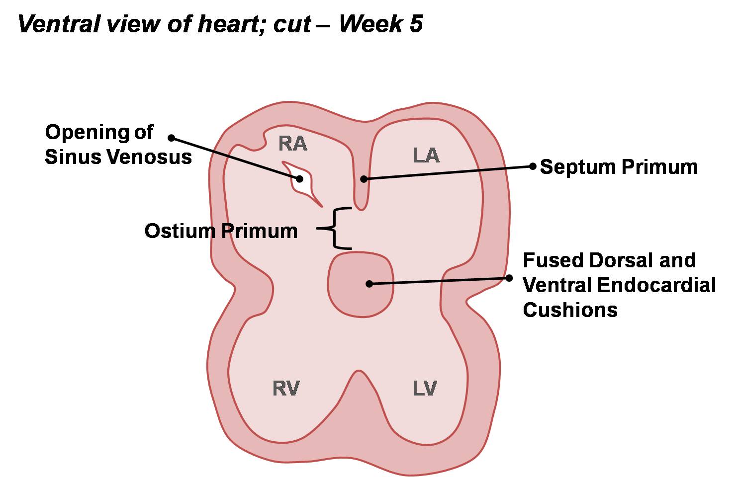
| 10:19, 14 March 2010 | Atrial Septation.jpg (file) |  |
90 KB | Z3212774 | category:Heart ILP During septation, the septum primum develops in the roof of the atrium, forming the early left and right atria. The opening of the sinus venosus has shifted to be incorporated into the right atrium. | 1 |
| 10:18, 14 March 2010 | AV Canal Division.jpg (file) |  |
93 KB | Z3212774 | category:Heart ILP Division of the atrioventricular canal occurs via growth and fusion of the dorsal and ventral (or superior and inferior) endocardial cushions. | 1 |
| 10:10, 14 March 2010 | Divisions of Early Heart Tube.jpg (file) |  |
85 KB | Z3212774 | {{Template:SEM}} category:Heart ILP | 1 |
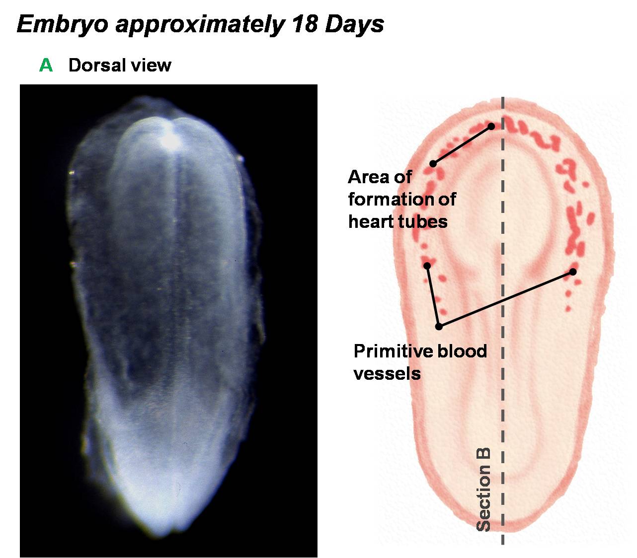
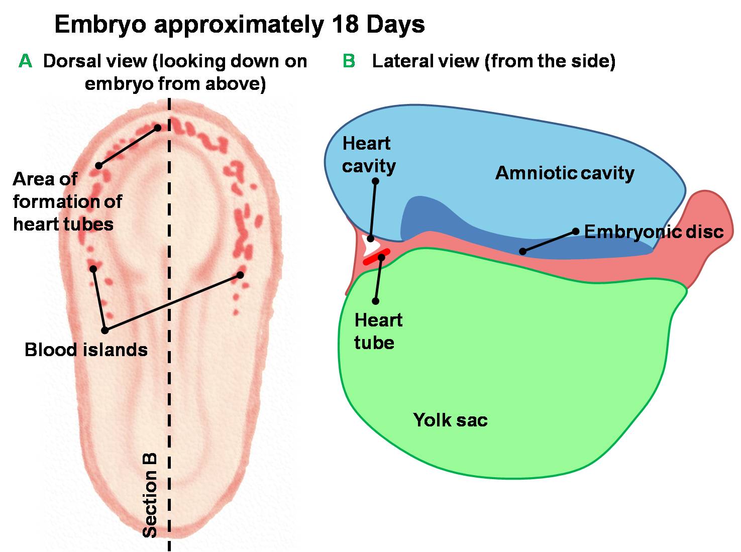
| 10:03, 14 March 2010 | Early Development of Heart Tube.jpg (file) |  |
132 KB | Z3212774 | Dorsal and lateral views of the earliest stages of cardiac development in the human embryo. Angiogenesis creates blood islands throughout the embryo during the third week of development. Angioblastic cords form in the cardiogenic mesoderm and canalise to | 1 |
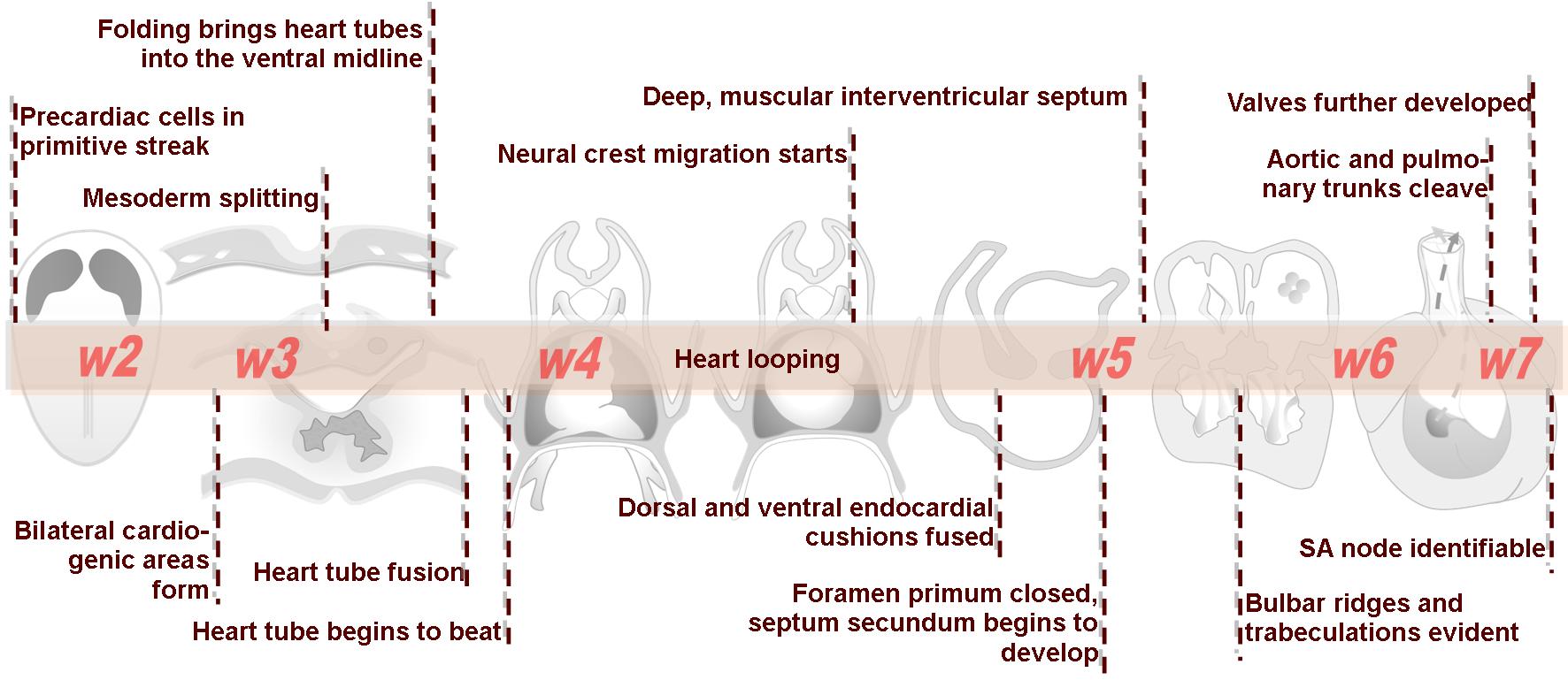
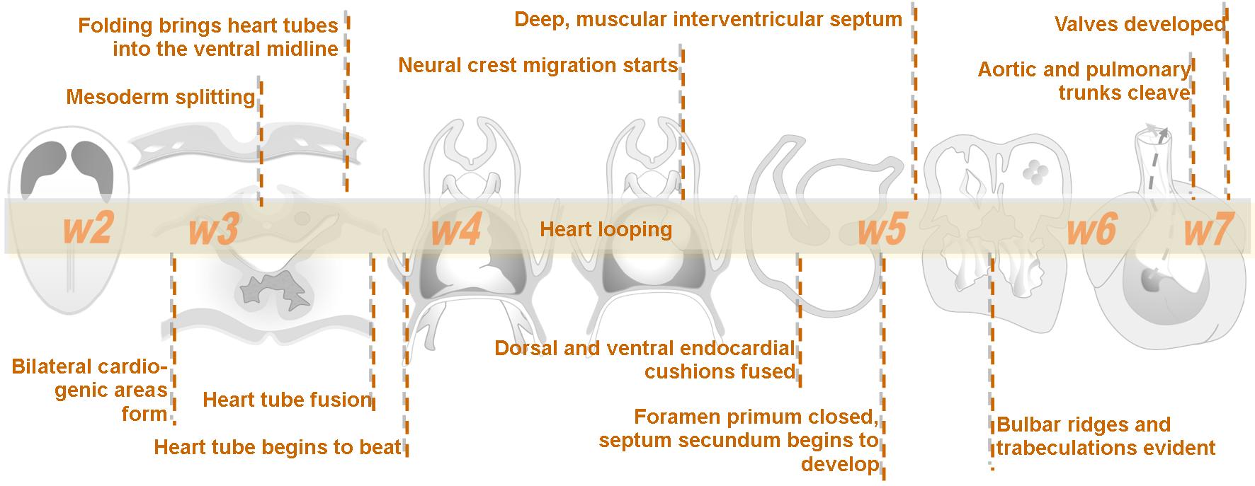
| 09:57, 14 March 2010 | Advanced Heart Development Timeline.jpg (file) |  |
158 KB | Z3212774 | category:Heart ILP | 1 |
| 09:56, 14 March 2010 | Intermediate Heart Development Timeline.jpg (file) |  |
133 KB | Z3212774 | category:Heart ILP | 1 |
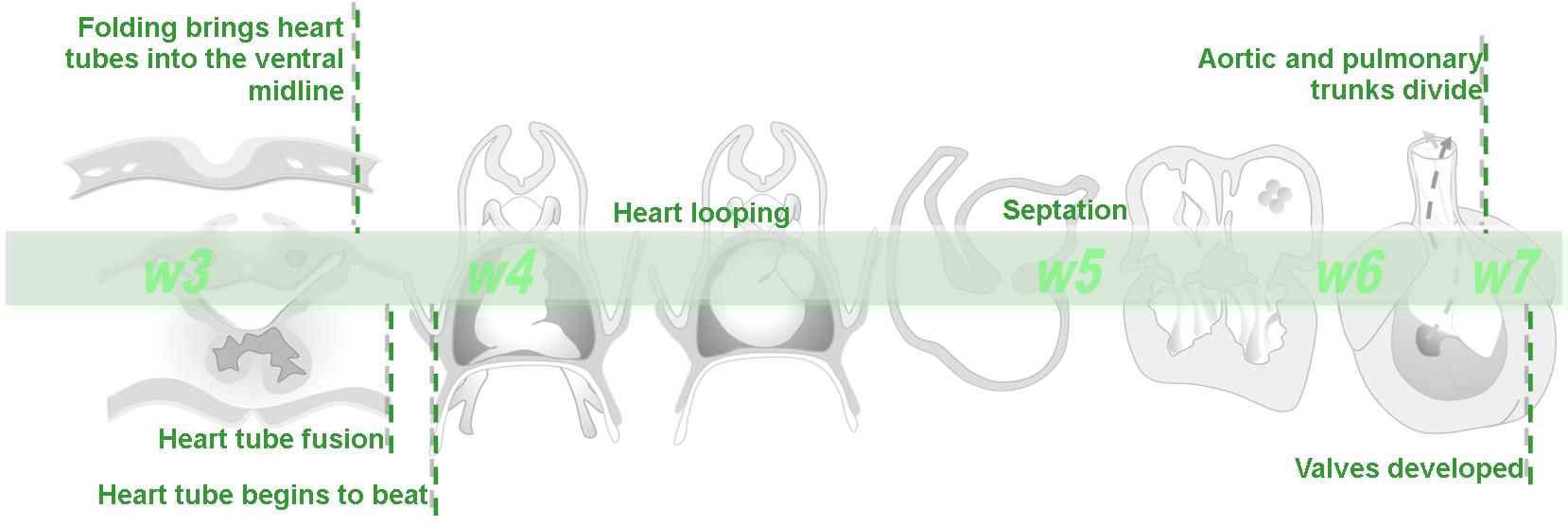
| 09:55, 14 March 2010 | Basic Heart Development Timeline.jpg (file) |  |
73 KB | Z3212774 | category:Heart ILP | 1 |
| 09:53, 14 March 2010 | Navigation bar 2.jpg (file) | 26 KB | Z3212774 | Heart ILP | 1 | |
| 09:52, 14 March 2010 | Navigation bar 1.jpg (file) | 31 KB | Z3212774 | Heart ILP | 1 | |
| 09:42, 14 March 2010 | Cardiac Embryology ILP Watermark.jpg (file) |  |
78 KB | Z3212774 | category:Heart ILP | 1 |
| 08:38, 14 March 2010 | Human Y chromosome.jpg (file) |  |
9 KB | S8600021 | 1 | |
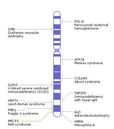
| 08:18, 14 March 2010 | Human X chromosome.jpg (file) |  |
25 KB | S8600021 | 1 | |
| 10:43, 12 March 2010 | Foundmedium.jpg (file) |  |
27 KB | S8600021 | 1 | |
| 10:42, 12 March 2010 | Foundsmall.jpg (file) |  |
11 KB | S8600021 | 1 | |
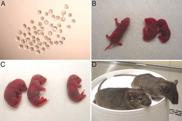
| 14:20, 24 February 2010 | Mice cloned from adult keratinocytes.jpg (file) |  |
20 KB | S8600021 | Mice cloned from adult skin keratinocytes and other cells by NT. (A) Cloned blastocysts from oocytes reconstructed by nuclear transfer (NT) of bulge follicle keratinocyte nuclei into unfertilized mouse oocytes. The α6+CD34+ bulge keratinocytes were iso | 1 |
| 11:14, 12 February 2010 | Citeulike 32x32.png (file) |  |
331 bytes | S8600021 | 1 | |
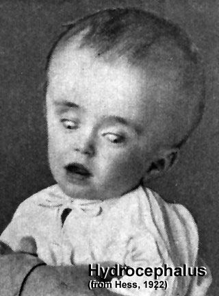
| 11:56, 26 January 2010 | Hydrocephalus.jpg (file) |  |
29 KB | S8600021 | Hydrocephalus (historic image from Hess, 1922) This is a defect of cerebrospinal fliud (CSF) flow, leading to enlarged ventricles and head, separated skull cranial sutures and fontanelles. Obstruction of CSF flow can occur at any time (prenatally or post | 1 |
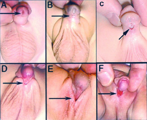
| 11:23, 26 January 2010 | Hypospadia classifications.jpg (file) |  |
47 KB | S8600021 | Classification of Hypospadias Hypospadia is classified by the location of the opening (meatus). A Anterior - on inferior surface of glans penis. B Coronal - in balanopenile furrow. C Distal - on distal third of shaft. D Penoscrotal - at base of shaf | 1 |
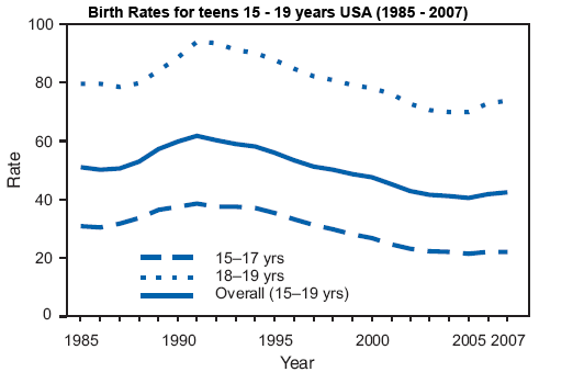
| 11:12, 26 January 2010 | Teen pregnancy USA.png (file) |  |
7 KB | S8600021 | Birth Rates* for Teens Aged 15 -19 Years, by Age Group United States, 1985 - 2007† * Per 1,000 women for specified age group. † Birth statistics are based on birth certificates filed in state vital statistics offices and reported to the National Ce | 1 |
| 10:54, 26 January 2010 | Aus multiple birth graph.png (file) |  |
5 KB | S8600021 | Australian pregnancies resulting in multiple births Multiple Births 1980 - 1.0% (2,249 of 223,318; 2,219 twins, 30 triplets or higher) 1990 - 1.2% (3,168 of 259,435; 3,074 twins, 94 triplets or higher) 2000 - 1.6% (3,900 of 245,700; 3,800 twins, 100 t | 1 |
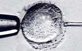
| 10:30, 26 January 2010 | In vitro fertilzation.jpg (file) |  |
4 KB | S8600021 | 1 | |
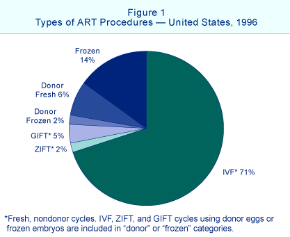
| 10:26, 26 January 2010 | ART types USA 1996.png (file) |  |
25 KB | S8600021 | Types of ART procedures USA 1996 Centre for Disease Control (USA) 1999 Survey of Assisted Reproductive Technology: Embryo Laboratory procedures and Practices (January 29, 1999) [http://embryology.med.unsw.edu.au/embryo/pdf/ARTsurvey.pdf PDF Version 473KB | 1 |
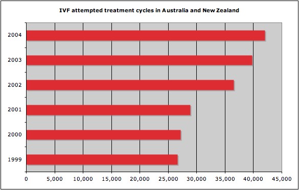
| 10:19, 26 January 2010 | IVF cycles ANZ 1999-2004.jpg (file) |  |
33 KB | S8600021 | IVF treatment cycles Australia and New Zealand 1999-2004. 2005 - 51,017 treatment cycles reported to ANZARD in Australia and New Zealand in 2005. Of these cycles, 91.1% were from Australian fertility centres and 8.9% from New Zealand’s centres. There i | 1 |
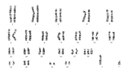
| 09:51, 26 January 2010 | Trisomy18male.jpg (file) |  |
10 KB | S8600021 | Trisomy 18 (Edwards Syndrome) karyotype. First recognized as a specific clinical entity by the discovery of an extra chromosome 18 in babies with a particular pattern of malformation by three independent groups (Edwards et al., Patau et al., Smith et al. | 1 |
| 14:05, 25 January 2010 | Drug-clearance-rates.png (file) |  |
14 KB | S8600021 | Infant Drug clearance rates The drug clearance data below are only approximate calculated rates for the fetus and infant from NZ Drug Safety in Lactation http://www.medsafe.govt.nz/Profs/PUarticles/lactation.htm#Infants | 1 |
| 13:22, 25 January 2010 | Perinatal mortality rate NSW 1992-2002.png (file) |  |
6 KB | S8600021 | Perinatal mortality rate NSW 1992-2002 http://embryology.med.unsw.edu.au/Child/birth7.htm NSW Statistics (Graph: Report of the New South Wales Chief Health Officer, 2004, http://www.health.nsw.gov.au/0 | 1 |
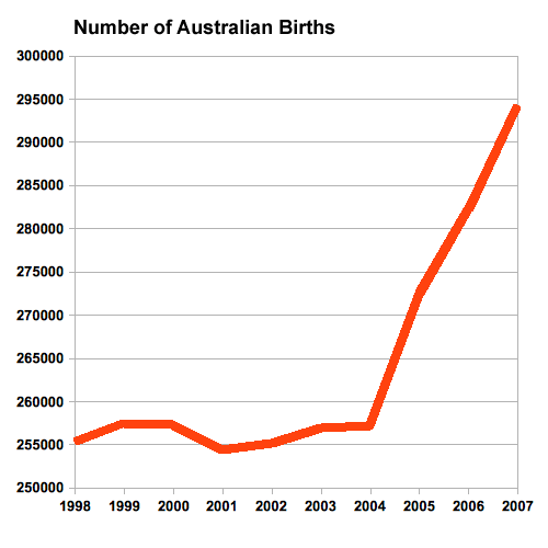
| 12:02, 25 January 2010 | Australian-births 2007.png (file) |  |
7 KB | S8600021 | Australian Births by Year Laws P & Sullivan EA 2009. Australia’s mothers and babies 2007. Perinatal statistics series no. 23. Cat. no. PER 48. Sydney: AIHW National Perinatal Statistics Unit. http://www.aihw.gov.au/publications/index.cfm/title/10972 | 1 |