File:Lymph node structure 02.jpg: Difference between revisions
(==Lymph node structure== Schematic representation of the organization of a lymph node (left panel). Afferent lymphatics enter lymph nodes and deliver lymph to the subcapsular sinus (SCS), which forms a channel around the periphery of the lymph node. Lymp) |
No edit summary |
||
| Line 11: | Line 11: | ||
[http://www.microbiol.unimelb.edu.au/research/immunology/s_mueller.html Mueller] | [http://www.microbiol.unimelb.edu.au/research/immunology/s_mueller.html Mueller] | ||
[[File_talk: | [[File_talk:Lymph_node_structure_02.jpg|Permissions]] | ||
Revision as of 18:54, 22 February 2012
Lymph node structure
Schematic representation of the organization of a lymph node (left panel). Afferent lymphatics enter lymph nodes and deliver lymph to the subcapsular sinus (SCS), which forms a channel around the periphery of the lymph node. Lymphatic sinuses run from the SCS through the cortex to the medulla, and exit the lymph node via efferent lymphatic vessels on the opposite, hilar, side of the organ. B cell follicles containing follicular dendritic cell (FDC) networks are arranged in the lymph node cortex and are separated from the SCS by a layer of marginal reticular cells (MRC). The T cells zones in the paracortex, which contain many fibroblastic reticular cells (FRC), are separated by the cortical ridge, an area rich in T cells, dendritic cells (DCs), blood vessels, and FRC. Blood vessels enter and exit the lymph node on the hilar side, and snake through the lymph node like the branches of a tree. Specialized high endothelial venules (HEVs) in the paracortex and cortical ridge allow entry of leukocytes from the blood. An image of a mouse popliteal lymph node generated using multi-colour immunofluoresence microscopy illustrates the distribution of CD3+ T cells (white), B220+ B cells (blue), CD11c+ DCs (green), LYVE1+ (Lymphatic Vessel Endothelial Receptor 1) lymphatics (cyan), PNAd+ (peripheral node addressin) HEVs (yellow), and ER-TR7+ stromal cells (red) (centre panel). The organization of stromal cells in the lymph node is shown by single-colour immunofluoresence staining for ER-TR7 (right panel).
Scale bars represent 200 μM.
Reference
<pubmed>19644499</pubmed>| PMC2785037 | Nat Rev Immunol.
File history
Click on a date/time to view the file as it appeared at that time.
| Date/Time | Thumbnail | Dimensions | User | Comment | |
|---|---|---|---|---|---|
| current | 18:53, 22 February 2012 | 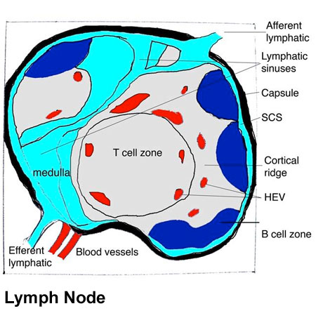 | 460 × 463 (54 KB) | Z8600021 (talk | contribs) | ==Lymph node structure== Schematic representation of the organization of a lymph node (left panel). Afferent lymphatics enter lymph nodes and deliver lymph to the subcapsular sinus (SCS), which forms a channel around the periphery of the lymph node. Lymp |
You cannot overwrite this file.
File usage
The following 4 pages use this file: