File:Ossification endochondral 1.jpg: Difference between revisions
From Embryology
No edit summary |
No edit summary |
||
| Line 1: | Line 1: | ||
Endochondral Ossification | '''Endochondral Ossification''' | ||
Histological image of a developing vertebra and intervertebral disc (rat) labelled. | |||
intervertebral disc - nucleus pulposus and annular fibrocartilage (bottom of image) | |||
vertebra - cartilage template and developing bony collar (top of image) | |||
scale bar 80 microns | |||
== Image version links == | == Image version links == | ||
| Line 6: | Line 14: | ||
[[:File:Ossification endochondral 1b.jpg|Medium 600px]] | [[:File:Ossification endochondral 1c.jpg|Small 400px]] | [[:File:Ossification endochondral 1b.jpg|Medium 600px]] | [[:File:Ossification endochondral 1c.jpg|Small 400px]] | ||
Original File Name: Endochondral9x10n3-1000px.jpg | |||
Image Source: UNSW Embryology | |||
[[Category:Histology]] [[Category:Musculoskeletal]] | [[Category:Histology]] [[Category:Musculoskeletal]] | ||
Revision as of 17:39, 15 September 2009
Endochondral Ossification
Histological image of a developing vertebra and intervertebral disc (rat) labelled.
intervertebral disc - nucleus pulposus and annular fibrocartilage (bottom of image)
vertebra - cartilage template and developing bony collar (top of image)
scale bar 80 microns
Image version links
Large 1000px | 800px | Medium 600px | Small 400px
Original File Name: Endochondral9x10n3-1000px.jpg
Image Source: UNSW Embryology
File history
Click on a date/time to view the file as it appeared at that time.
| Date/Time | Thumbnail | Dimensions | User | Comment | |
|---|---|---|---|---|---|
| current | 10:53, 14 September 2009 | 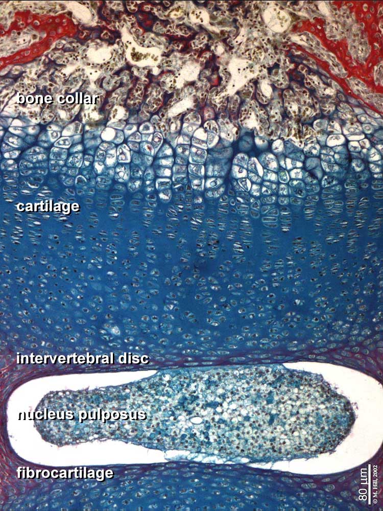 | 750 × 1,000 (147 KB) | S8600021 (talk | contribs) | Endochondral9x10n3-1000px.jpg |
You cannot overwrite this file.
File usage
The following page uses this file: