File:Human right ovary and tube 1.jpg: Difference between revisions
From Embryology
No edit summary |
|||
| Line 5: | Line 5: | ||
Note the relative size and position of the ovary with respect to the uterine tube (fallopian tube). The ovary appears white and relatively avascular on its surface. In the background the associated mesenteries and peritoneal cavity can be seen. | Note the relative size and position of the ovary with respect to the uterine tube (fallopian tube). The ovary appears white and relatively avascular on its surface. In the background the associated mesenteries and peritoneal cavity can be seen. | ||
''' | |||
'''Links:''' [[Ovary Development]] | [[Uterus Development]] | [[Menstrual Cycle]] | [[:File:Human_ovulation_01.jpg|Ovulation Image]] | |||
Revision as of 06:56, 8 December 2010
Human Ovary and Associated Uterine Tube
Adult human ovary (right) viewed by laparoscopy.
Note the relative size and position of the ovary with respect to the uterine tube (fallopian tube). The ovary appears white and relatively avascular on its surface. In the background the associated mesenteries and peritoneal cavity can be seen.
Links: Ovary Development | Uterus Development | Menstrual Cycle | Ovulation Image
Image source: UNSW Embryology, no reproduction without permission.
File history
Yi efo/eka'e gwa ebo wo le nyangagi wuncin ye kamina wunga tinya nan
| Gwalagizhi | Nyangagi | Dimensions | User | Comment | |
|---|---|---|---|---|---|
| current | 09:31, 9 April 2010 | 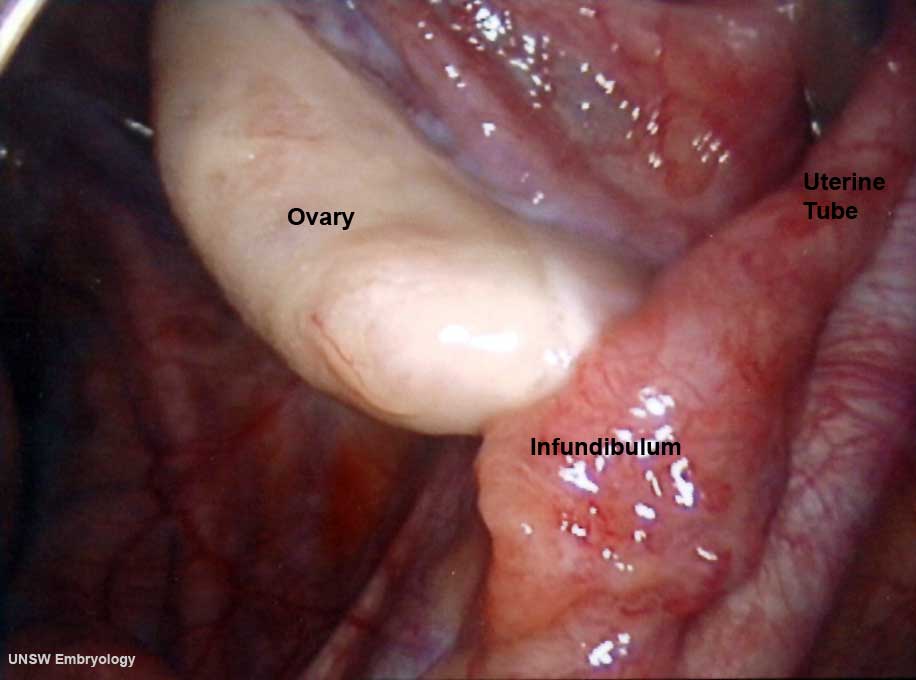 | 916 × 680 (32 KB) | S8600021 (talk | contribs) | Human right ovary and tube as viewed by laparoscopy == Image version links == Large 1000px | 800px | Medium 600px | [[:F |
You cannot overwrite this file.