File:Plasma cell EM06.jpg: Difference between revisions
No edit summary |
m (→Reference) |
||
| Line 19: | Line 19: | ||
===Reference=== | ===Reference=== | ||
{{#pmid:4563148}} | |||
Revision as of 16:11, 17 February 2019
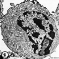
Plasma Cell Electron Micrograph
- Plasma cell (effector B cell) is the terminally differentiated B lymphocyte.
- increase in rough endoplasmic reticulum (RER) associated with antibody production.
Fig 6. Plasma cells labeled by incubations with aMBLA, followed by the "bridge"technique with SaRIg-Pox.
Plasma cells, especially when fully mature, showed also a reduced amount of MBLA compared with B lymphocytes.
MBLA - mouse-specific bone marrow-derived lymphocyte antigens, label for B cells.
X 7500.
- Lymphocyte EM Images: T and B Lymphocytes 1 TEM | T and B Lymphocytes 2 TEM | T Lymphocyte SEM | B lymphocyte 1 TEM | B lymphocyte 2 TEM | B lymphocyte 3 TEM | Plasma Cell TEM | T2 Lymphocyte 1 TEM | T2 Lymphocyte 2 TEM | lymphocyte rosettes | T lymphocyte 1 | T lymphocyte 2 | T lymphocyte 3 | T lymphocyte 4 | T lymphocyte 5 | T lymphocyte 6 | B lymphocyte | B lymphocytes TEM | Immune System Development | Blood
Reference
Matter A, Lisowska-Bernstein B, Ryser JE, Lamelin JP & Vassalli P. (1972). Mouse thymus-independent and thymus-derived lymphoid cells. II. Ultrastructural studies. J. Exp. Med. , 136, 1008-30. PMID: 4563148
Copyright
Rockefeller University Press - Copyright Policy This article is distributed under the terms of an Attribution–Noncommercial–Share Alike–No Mirror Sites license for the first six months after the publication date (see http://www.jcb.org/misc/terms.shtml). After six months it is available under a Creative Commons License (Attribution–Noncommercial–Share Alike 4.0 Unported license, as described at https://creativecommons.org/licenses/by-nc-sa/4.0/ ). (More? Help:Copyright Tutorial)
File history
Yi efo/eka'e gwa ebo wo le nyangagi wuncin ye kamina wunga tinya nan
| Gwalagizhi | Nyangagi | Dimensions | User | Comment | |
|---|---|---|---|---|---|
| current | 15:49, 22 February 2012 |  | 595 × 600 (84 KB) | Z8600021 (talk | contribs) | ==Plasma Cell Electron Micrograph== Fig 6. Plasma cells labeled by incubations with aMBLA, followed by the "bridge"technique with SaRIg-Pox. Plasma cells, especially when fully mature, showed also a reduced amount of MBLA compared with B lymphocytes. |
You cannot overwrite this file.
File usage
The following 9 pages use this file:
- ANAT2241 Lymphatic Tissue and Immune System
- Immune System - Antibody Development
- Immune System Development
- Lymph Node Development
- SH Lecture - Lymphatic Structure and Organs
- SH Practical - Lymphatic Quiz
- SH Practical - Lymphatic Structure and Organs
- Talk:SH Practical - Lymphatic Quiz
- Template talk:Lymphocyte images