2018 Group Project 4: Difference between revisions
No edit summary |
|||
| Line 8: | Line 8: | ||
==Cardiac neural crest cells== | ==Cardiac neural crest cells== | ||
Neural crest cells are a population of multipotent cells which arises during embryonic development at the dorsal neural tube. These cells are capable of migrating and differentiating throughout the body to give rise to many different cell types. The cardiac neural crest cells (CNCCs) are a subpopulation of the cranial neural crest cells and migrate ventrally from the dorsal neural tube and accumulate in the circumpharyngeal ridge. {{#pmid:PMC4288758|PMC4288758}} The cardiac neural crest wells will then proceed into the pharyngeal arches as each arch develops. | |||
CNCCs are not critical in the initial formation of vessels but they are needed for the remodelling of subsequent arteries, and will give rise to the smooth muscle tunics of the great arteries.{{#pmid:PMC4288758|PMC4288758}} | |||
==History of cardiac neural crest cells== | ==History of cardiac neural crest cells== | ||
Revision as of 09:15, 8 September 2018
| Projects 2018: 1 Adrenal Medulla | 3 Melanocytes | 4 Cardiac | 5 Dorsal Root Ganglion |
Project Pages are currently being updated (notice removed when completed)
Neural Crest and Cardiac Development
Introduction of the heart
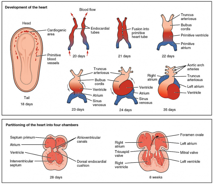
The heart is a muscular organ which plays a critical role in the circulatory system by mechanically pumping blood to various organs around the body for the exchange of nutrients and gases. It is located at the center of the chest, right behind the sternum and is tilted slightly to the left. The heart has four different chambers which are compartmentalized by semilunar and atrioventricular valves into the left and right atria and ventricles
Cardiac neural crest cells
Neural crest cells are a population of multipotent cells which arises during embryonic development at the dorsal neural tube. These cells are capable of migrating and differentiating throughout the body to give rise to many different cell types. The cardiac neural crest cells (CNCCs) are a subpopulation of the cranial neural crest cells and migrate ventrally from the dorsal neural tube and accumulate in the circumpharyngeal ridge. PubmedParser error: Invalid PMID, please check. (PMID: [1]) The cardiac neural crest wells will then proceed into the pharyngeal arches as each arch develops.
CNCCs are not critical in the initial formation of vessels but they are needed for the remodelling of subsequent arteries, and will give rise to the smooth muscle tunics of the great arteries.PubmedParser error: Invalid PMID, please check. (PMID: [2])
History of cardiac neural crest cells
"In a chick study of parasympathetic innervation of the heart, Margaret Kirby and colleagues ablated neural crest and serendipitously discovered that the embryos lacked aorticopulmonary septation (Kirby et al., 1983). The subregion of cranial neural crest ablated by Dr. Kirby has been called the “cardiac neural crest”, not because the cells of this region migrate solely to the heart, but for the importance of crest-derived ectomesenchyme in cardiovascular development. "
