File:Gilmour1941 plate07.jpg: Difference between revisions
mNo edit summary |
mNo edit summary |
||
| Line 5: | Line 5: | ||
[[:File:Gilmour1941 fig02.jpg|'''Fig. 2.''']] 125 mm foetus. Focus of megalo-normoblaetic erythropoiesis in neck. One early and several intermediate megaloblasts forming pyknotic megaloblasts and - with loss of nucleus - a Inegalpcyte, and — from shrinkage in size — orthochrematic normoblasts and — by loss of nucleus - a normocyte. Jenner. | [[:File:Gilmour1941 fig02.jpg|'''Fig. 2.''']] 125 mm foetus. Focus of megalo-normoblaetic erythropoiesis in neck. One early and several intermediate megaloblasts forming pyknotic megaloblasts and - with loss of nucleus - a Inegalpcyte, and — from shrinkage in size — orthochrematic normoblasts and — by loss of nucleus - a normocyte. Jenner. | ||
{{Online Editor}} - Note this is an historic representation of haemopoiesis that may differ from our current understanding of {{blood}} development. | |||
{{Gilmour1941 figures}} | {{Gilmour1941 figures}} | ||
Revision as of 11:11, 17 May 2018
Plate VII
Fig. 1. Red blood cells and their precursors. 1 and 2, haemocytoblasts ; 3, 4 and 5, early, intermediate and late primary erythroblasts; 6 and 7, basophil and erthochromatic normoblasts; 8, normocyte; 9, 10, l1 and 12, early, intermediate, late and pyknotic megaloblasts; 13, megalocyte ; 14, 15, 16 and 17 early, intermediate, late and pyknotic primitive megaloblasts; 18, primitive erythrocyte; 4a, basophil normoblast with prematurely pyknotic nucleus; 10a, megaloblast with prematurely pyknotic nueleus. Jenner.
Fig. 2. 125 mm foetus. Focus of megalo-normoblaetic erythropoiesis in neck. One early and several intermediate megaloblasts forming pyknotic megaloblasts and - with loss of nucleus - a Inegalpcyte, and — from shrinkage in size — orthochrematic normoblasts and — by loss of nucleus - a normocyte. Jenner.
Online Editor - Note this is an historic representation of haemopoiesis that may differ from our current understanding of blood development.
| Historic Disclaimer - information about historic embryology pages |
|---|
 |
Figure Links: Plate 7 | Fig. 1 | Fig. 2 | Plate 8 | Fig. 3 | Fig. 4 | Fig. 5 | Plate 9 | Fig. 6 | Fig. 8 | Plate 10 | Fig. 7 | Fig. 9 |Fig. 10 | Fig. 11 | Gilmour 1941 | Modern notes - blood | Hematopoietic and stromal cell differentiation
Reference
Gilmour JR. Normal haemopoiesis in intra-uterine and neonatal life. (1941) J. Pathol. Bacteriol. 52: 25-55.
Cite this page: Hill, M.A. (2024, June 14) Embryology Gilmour1941 plate07.jpg. Retrieved from https://embryology.med.unsw.edu.au/embryology/index.php/File:Gilmour1941_plate07.jpg
- © Dr Mark Hill 2024, UNSW Embryology ISBN: 978 0 7334 2609 4 - UNSW CRICOS Provider Code No. 00098G
File history
Click on a date/time to view the file as it appeared at that time.
| Date/Time | Thumbnail | Dimensions | User | Comment | |
|---|---|---|---|---|---|
| current | 01:09, 17 May 2018 | 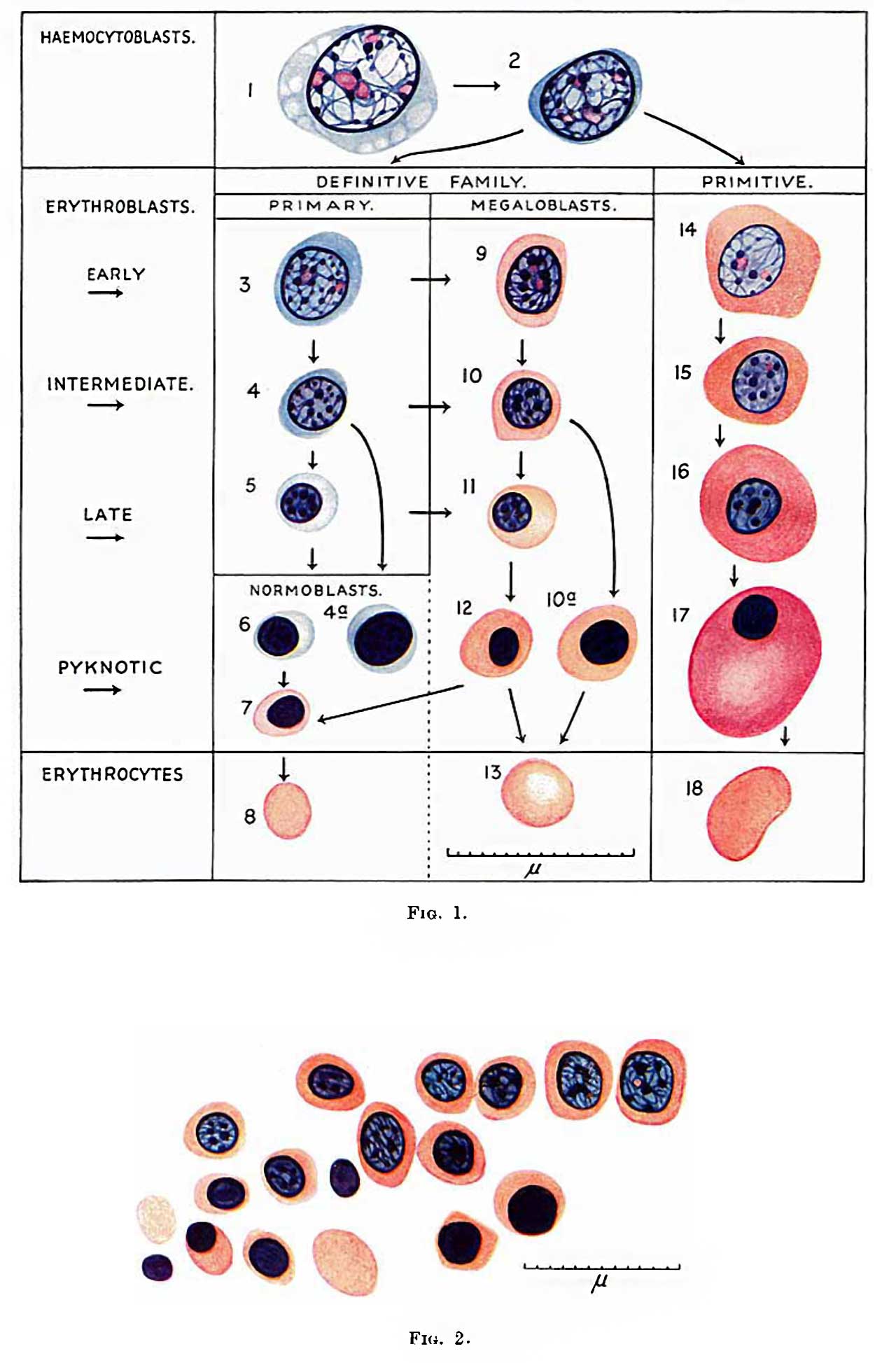 | 1,280 × 1,947 (160 KB) | Z8600021 (talk | contribs) | |
| 01:08, 17 May 2018 |  | 1,606 × 2,605 (235 KB) | Z8600021 (talk | contribs) | {{Ref-Gilmour1941}} |
You cannot overwrite this file.
File usage
The following page uses this file: