File:Gilmour1941 plate09.jpg: Difference between revisions
mNo edit summary |
(Z8600021 uploaded a new version of File:Gilmour1941 plate09.jpg) |
(No difference)
| |
Revision as of 10:22, 17 May 2018
Plate IX
Fig. 6. Embryo 4, Frazer, about 20 pairs of somites. Blood vessels in yolk sac full of primitive erythroblasts. (Stain - Haematoxylin Eosin) Dufay process. x550.
Fig. 8. 10 mm embryo (Frazer). Two haemocytoblasts among epithelial cells of yolk sac. Primitive erythroblasts in yolk sac vessel. Ehrlich’s haematoxylin and eosin. Dufay process. x1100.
Reference
Gilmour JR. Normal haemopoiesis in intra-uterine and neonatal life. (1941) J. Pathol. Bacteriol. 52: 25-55.
Cite this page: Hill, M.A. (2024, June 2) Embryology Gilmour1941 plate09.jpg. Retrieved from https://embryology.med.unsw.edu.au/embryology/index.php/File:Gilmour1941_plate09.jpg
- © Dr Mark Hill 2024, UNSW Embryology ISBN: 978 0 7334 2609 4 - UNSW CRICOS Provider Code No. 00098G
File history
Click on a date/time to view the file as it appeared at that time.
| Date/Time | Thumbnail | Dimensions | User | Comment | |
|---|---|---|---|---|---|
| current | 10:22, 17 May 2018 | 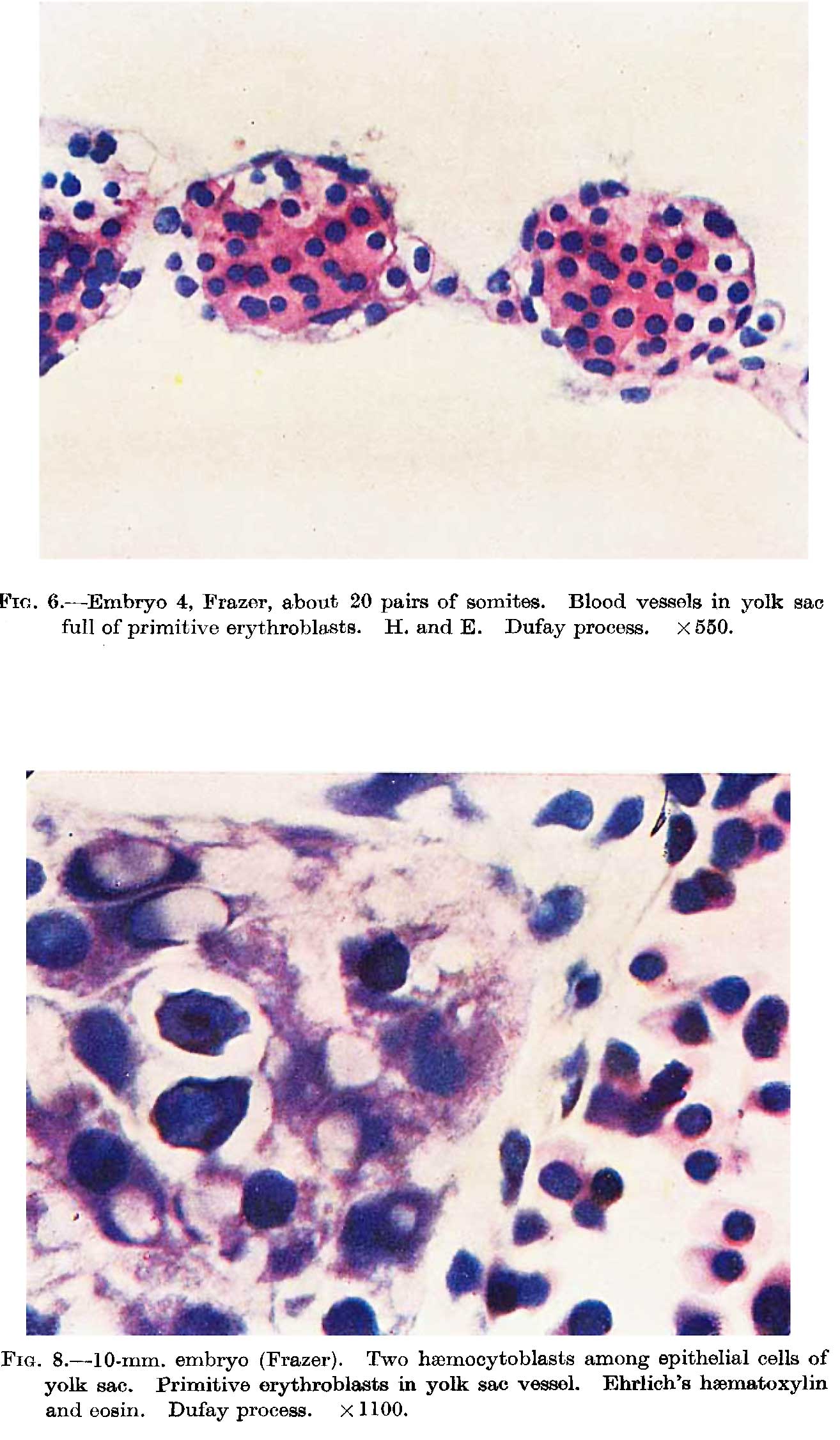 | 1,314 × 2,252 (212 KB) | Z8600021 (talk | contribs) | |
| 10:18, 17 May 2018 | 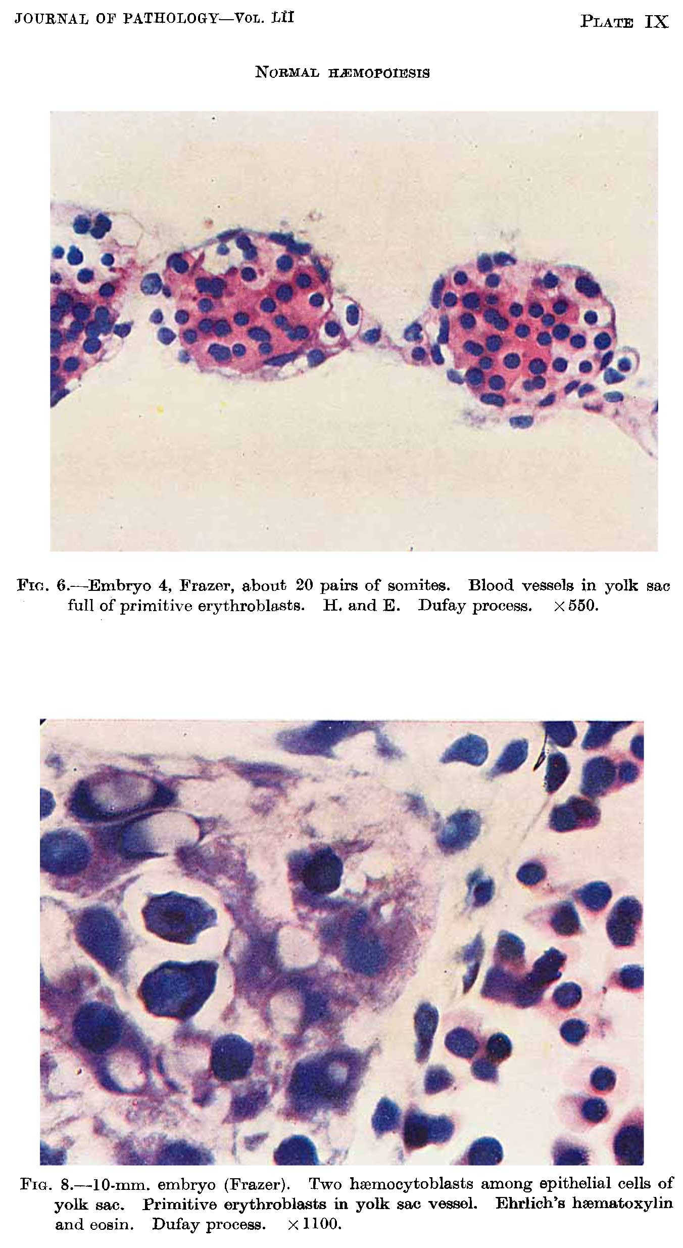 | 1,352 × 2,469 (247 KB) | Z8600021 (talk | contribs) |
You cannot overwrite this file.
File usage
The following page uses this file: