File:Gland duct histology cartoon.jpg: Difference between revisions
mNo edit summary |
|||
| Line 1: | Line 1: | ||
==Gland Duct Histology Cartoon== | ==Gland Duct Histology Cartoon== | ||
Cartoon shows the general organisation of salivary gland duct system. | Cartoon shows the general organisation of {{salivary gland}} duct system. | ||
| Line 19: | Line 19: | ||
:'''Parotid Gland Links:''' [[:File:Parotid gland histology 01.jpg|Gland overview]] | [[:File:Parotid gland histology 06.jpg|lobe overview]] | [[:File:Parotid gland histology 04.jpg|serous acini]] | [[:File:Parotid gland histology 02.jpg|Striated duct and serous]] | [[:File:Parotid gland histology 03.jpg|Intercalated duct]] | [[:File:Parotid gland histology 05.jpg|excretory duct]] | [[:File:Parotid_histology_stratified_columnar_01.jpg|Gland epithelium]] | [[:File:Gland duct histology cartoon.jpg|Duct cartoon]] | :'''Parotid Gland Links:''' [[:File:Parotid gland histology 01.jpg|Gland overview]] | [[:File:Parotid gland histology 06.jpg|lobe overview]] | [[:File:Parotid gland histology 04.jpg|serous acini]] | [[:File:Parotid gland histology 02.jpg|Striated duct and serous]] | [[:File:Parotid gland histology 03.jpg|Intercalated duct]] | [[:File:Parotid gland histology 05.jpg|excretory duct]] | [[:File:Parotid_histology_stratified_columnar_01.jpg|Gland epithelium]] | [[:File:Gland duct histology cartoon.jpg|Duct cartoon]] | ||
{{Footer}} | |||
[[Category:Gland]] | [[Category:Gland]] | ||
Latest revision as of 18:09, 16 April 2018
Gland Duct Histology Cartoon
Cartoon shows the general organisation of salivary gland duct system.
Interlobular and interlobar ducts - embedded in the connective tissue surrounding the lobes and lobules of the glands
stratified cuboidal or stratified columnar epithelium
stratified squamous epithelium at the oral cavity opening
Intralobular ducts - in between the secretory acini within the lobules
Intercalated ducts - difficult to identify in mucous glands
Striated ducts - absent in purely mucous glands
- Parotid Gland Links: Gland overview | lobe overview | serous acini | Striated duct and serous | Intercalated duct | excretory duct | Gland epithelium | Duct cartoon
Cite this page: Hill, M.A. (2024, June 22) Embryology Gland duct histology cartoon.jpg. Retrieved from https://embryology.med.unsw.edu.au/embryology/index.php/File:Gland_duct_histology_cartoon.jpg
- © Dr Mark Hill 2024, UNSW Embryology ISBN: 978 0 7334 2609 4 - UNSW CRICOS Provider Code No. 00098G
File history
Yi efo/eka'e gwa ebo wo le nyangagi wuncin ye kamina wunga tinya nan
| Gwalagizhi | Nyangagi | Dimensions | User | Comment | |
|---|---|---|---|---|---|
| current | 10:55, 26 March 2014 | 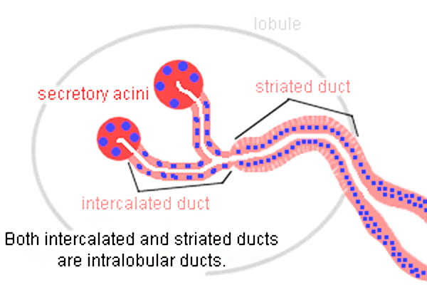 | 600 × 400 (41 KB) | Z8600021 (talk | contribs) |
You cannot overwrite this file.
File usage
The following 3 pages use this file: