File:Basu1932 fig01.jpg: Difference between revisions
From Embryology
(Z8600021 uploaded a new version of File:Basu1932 fig01.jpg) |
mNo edit summary |
||
| Line 1: | Line 1: | ||
The two drawings—one of the anterior and the other of the posterior view of the heart and the great blood vessels—illustrate the abnormalities of the specimen. | |||
===Reference=== | |||
{{Ref-Basu1932}} | |||
{{Footer}} | |||
Latest revision as of 09:06, 29 November 2017
The two drawings—one of the anterior and the other of the posterior view of the heart and the great blood vessels—illustrate the abnormalities of the specimen.
Reference
Basu BN Persistent left superior vena cava, left duct of cuvier and left horn of the sinus venosus. (1932) J Anat. 66(2): 268–270. PMID 17104374
Cite this page: Hill, M.A. (2024, June 26) Embryology Basu1932 fig01.jpg. Retrieved from https://embryology.med.unsw.edu.au/embryology/index.php/File:Basu1932_fig01.jpg
- © Dr Mark Hill 2024, UNSW Embryology ISBN: 978 0 7334 2609 4 - UNSW CRICOS Provider Code No. 00098G
File history
Yi efo/eka'e gwa ebo wo le nyangagi wuncin ye kamina wunga tinya nan
| Gwalagizhi | Nyangagi | Dimensions | User | Comment | |
|---|---|---|---|---|---|
| current | 09:05, 29 November 2017 | 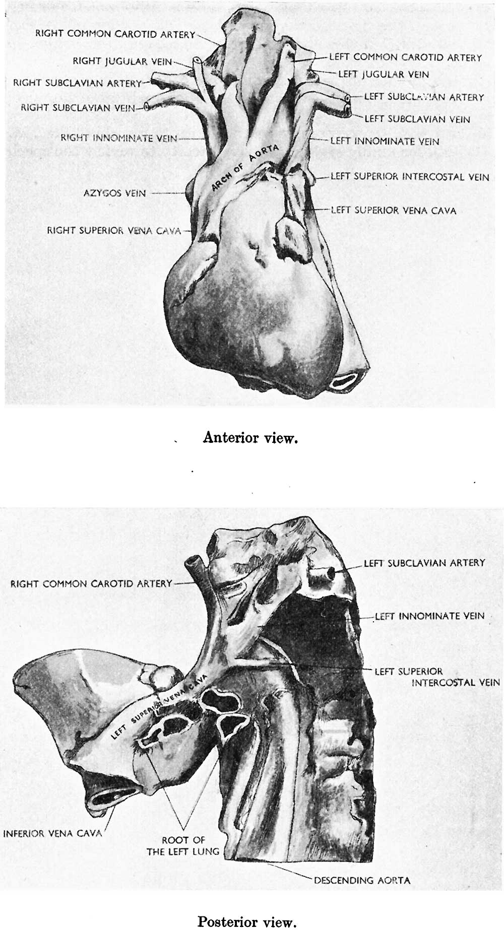 | 1,000 × 1,846 (190 KB) | Z8600021 (talk | contribs) | |
| 09:05, 29 November 2017 | 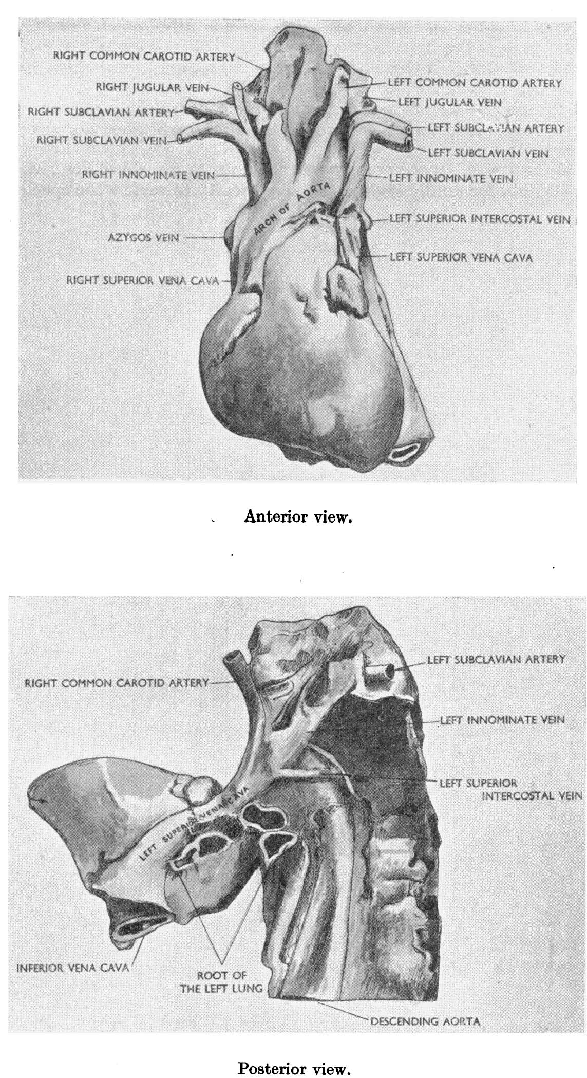 | 1,143 × 2,098 (406 KB) | Z8600021 (talk | contribs) |
You cannot overwrite this file.